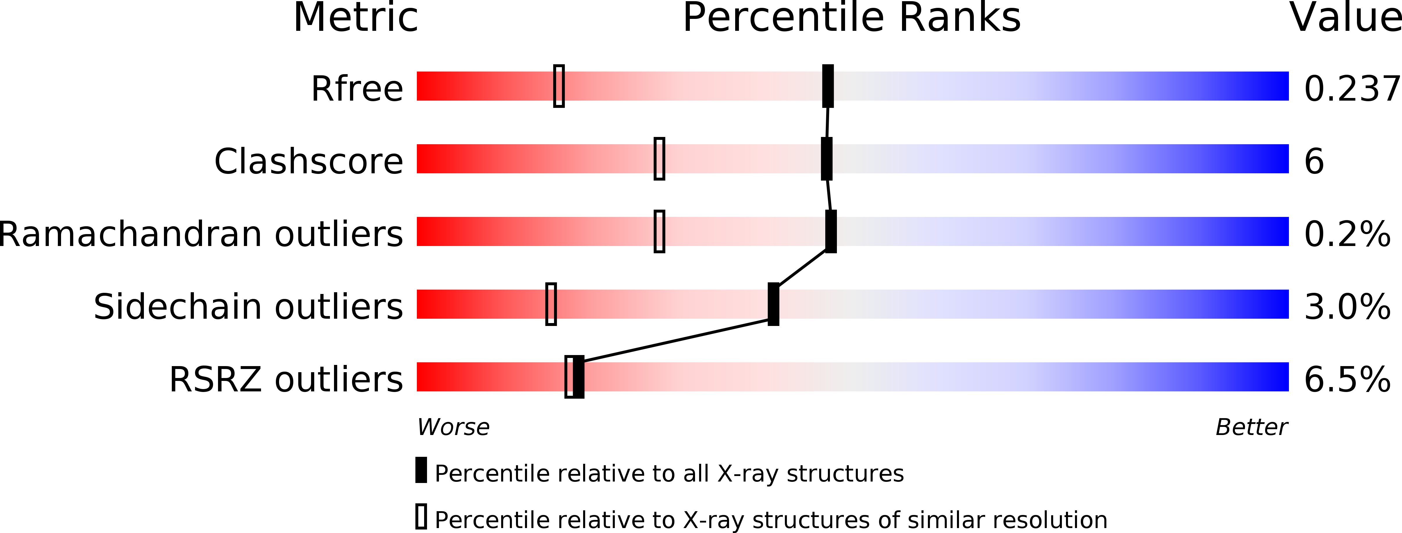
1W0D image
Deposition Date 2004-06-03
Release Date 2004-12-14
Last Version Date 2024-05-08
Entry Detail
PDB ID:
1W0D
Keywords:
Title:
The high resolution structure of Mycobacterium tuberculosis LeuB (Rv2995c)
Biological Source:
Source Organism:
Host Organism:
Method Details:
Experimental Method:
Resolution:
1.65 Å
R-Value Free:
0.24
R-Value Work:
0.21
R-Value Observed:
0.21
Space Group:
P 21 21 21
Macromolecular Entities
Structures with similar UniProt ID
Protein Blast
Polymer Type:polypeptide(L)
Molecule:3-ISOPROPYLMALATE DEHYDROGENASE
Chain IDs:A, B, C, D
Chain Length:337
Number of Molecules:4
Biological Source:MYCOBACTERIUM TUBERCULOSIS
Ligand Molecules
Primary Citation
The High Resolution Structure of Leub (Rv2995C) from Mycobacterium Tuberculosis
J.Mol.Biol. 346 1 ? (2005)
PMID: 15663922 DOI: 10.1016/J.JMB.2004.11.059

Abstact

The crystal structure of the enzyme 3-isopropylmalate dehydrogenase (IPMDH) from Mycobacterium tuberculosis (LeuB, Mtb-IPMDH, Rv2995c) without substrate or co-factor was determined at 1.65 A resolution, which is the highest resolution reported for an IPMDH to date. The crystals contain two functional dimers in the asymmetric unit in an arrangement close to a tetramer of D2 symmetry. Despite the absence of a substrate or inhibitor bound to the protein, the structure of the monomer resembles the previously observed closed form of the enzyme more closely than the open form. A comparison with the substrate complex of IPMDH from Thiobacillus ferrooxidans and the co-factor complex of the Thermus thermophilus enzyme revealed a close relationship of the active-site architecture between the various bacterial enzymes. The inhibitor O-isobutenyl oxalylhydroxamate was found to bind to the active site of IPMDH in a mode similar to the substrate isopropylmalate.

Legend

Protein

Chemical

Disease

Primary Citation of related structures