1W07 image
Deposition Date 2004-06-01
Release Date 2004-12-15
Last Version Date 2024-11-06
Entry Detail
PDB ID:
1W07
Keywords:
Title:
Arabidopsis thaliana acyl-CoA oxidase 1
Biological Source:
Source Organism:
Host Organism:
Method Details:
Experimental Method:
Resolution:
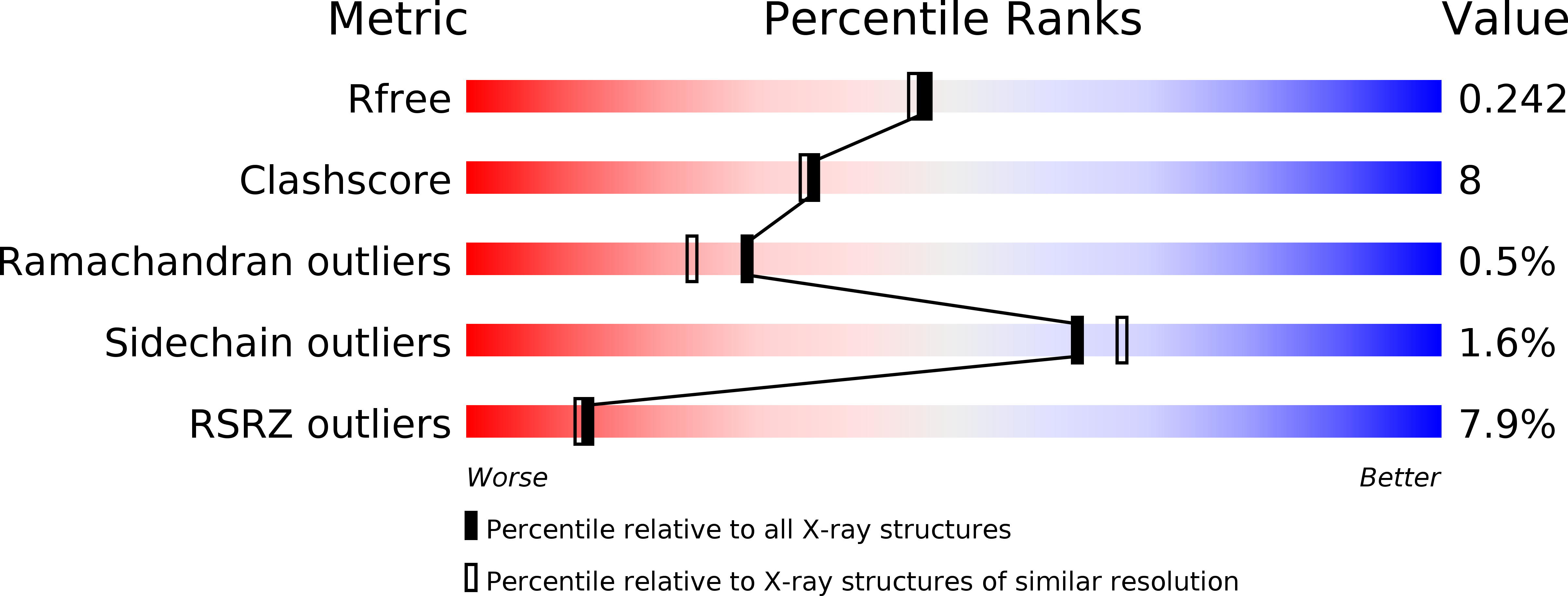
2.00 Å
R-Value Free:
0.24
R-Value Work:
0.20
R-Value Observed:
0.20
Space Group:
P 21 21 21
Macromolecular Entities
Structures with similar UniProt ID
Protein Blast
Polymer Type:polypeptide(L)
Molecule:ACYL-COA OXIDASE
Gene (Uniprot):ACX1
Mutagens:YES
Chain IDs:A, B
Chain Length:659
Number of Molecules:2
Biological Source:ARABIDOPSIS THALIANA
Primary Citation
Acyl-Coa Oxidase 1 from Arabidopsis Thaliana. Structure of a Key Enzyme in Plant Lipid Metabolism
J.Mol.Biol. 345 487 ? (2005)
PMID: 15581893 DOI: 10.1016/J.JMB.2004.10.062

Abstact

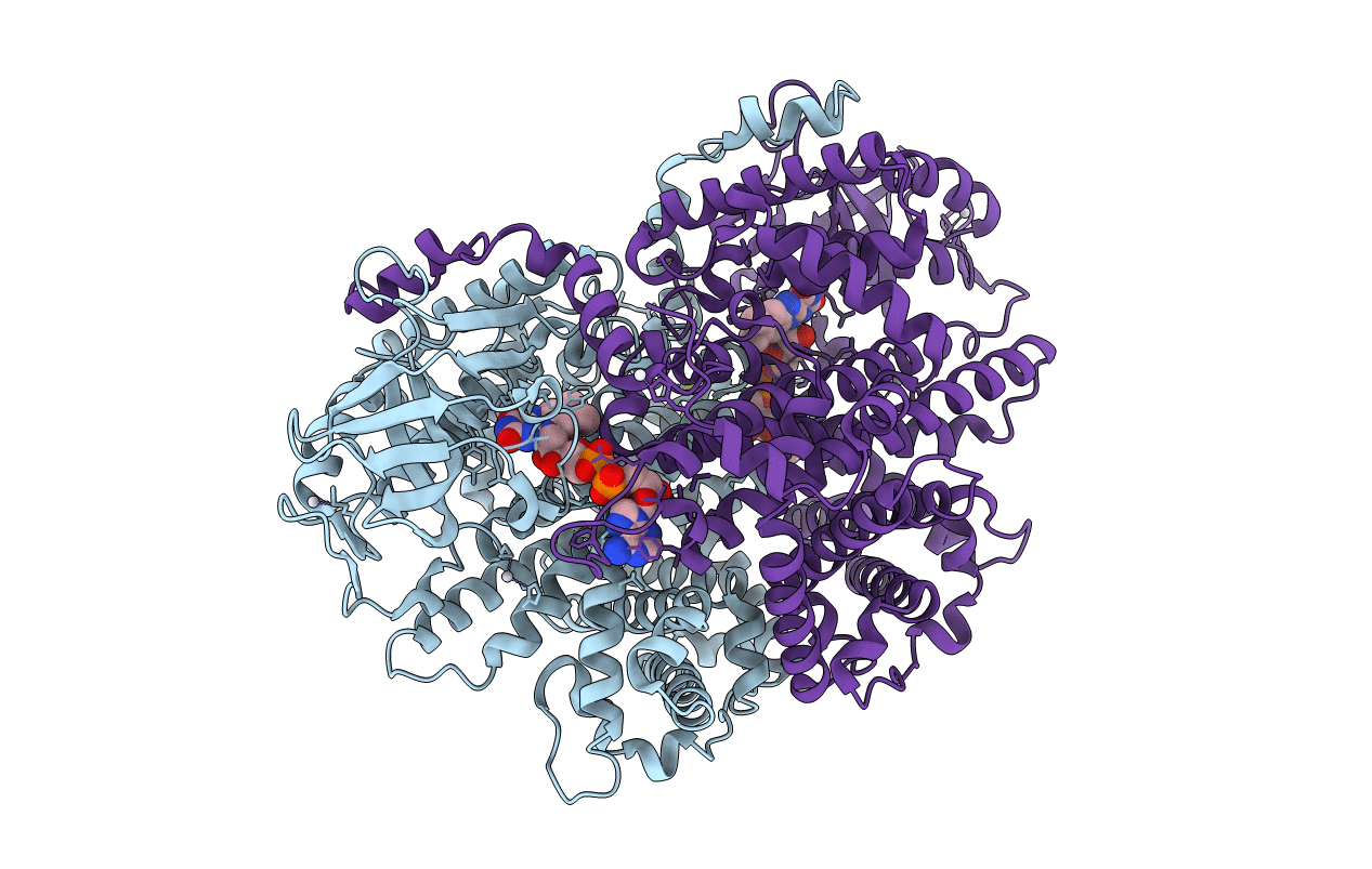
The peroxisomal acyl-CoA oxidase family plays an essential role in lipid metabolism by catalyzing the conversion of acyl-CoA into trans-2-enoyl-CoA during fatty acid beta-oxidation. Here, we report the X-ray structure of the FAD-containing Arabidopsis thaliana acyl-CoA oxidase 1 (ACX1), the first three-dimensional structure of a plant acyl-CoA oxidase. Like other acyl-CoA oxidases, the enzyme is a dimer and it has a fold resembling that of mammalian acyl-CoA oxidase. A comparative analysis including mammalian acyl-CoA oxidase and the related tetrameric mitochondrial acyl-CoA dehydrogenases reveals a substrate-binding architecture that explains the observed preference for long-chained, mono-unsaturated substrates in ACX1. Two anions are found at the ACX1 dimer interface and for the first time the presence of a disulfide bridge in a peroxisomal protein has been observed. The functional differences between the peroxisomal acyl-CoA oxidases and the mitochondrial acyl-CoA dehydrogenases are attributed to structural differences in the FAD environments.

Legend

Protein

Chemical

Disease

Primary Citation of related structures