
Deposition Date
2004-05-25
Release Date
2004-09-02
Last Version Date
2024-05-15
Entry Detail
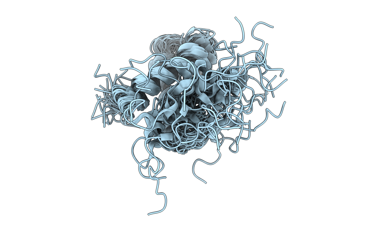
PDB ID:
1VZS
Keywords:
Title:
Solution structure of subunit F6 from the peripheral stalk region of ATP synthase from bovine heart mitochondria
Biological Source:
Source Organism(s):
BOS TAURUS (Taxon ID: 9913)
Expression System(s):
Method Details:
Experimental Method:
Conformers Calculated:
50
Conformers Submitted:
34
Selection Criteria:
JUMP IN TOTAL ENERGIES
