1VZO image
Deposition Date 2004-05-21
Release Date 2004-06-11
Last Version Date 2023-12-13
Entry Detail
PDB ID:
1VZO
Keywords:
Title:
The structure of the N-terminal kinase domain of MSK1 reveals a novel autoinhibitory conformation for a dual kinase protein
Biological Source:
Source Organism:
HOMO SAPIENS (Taxon ID: 9606)
Host Organism:
Method Details:
Experimental Method:
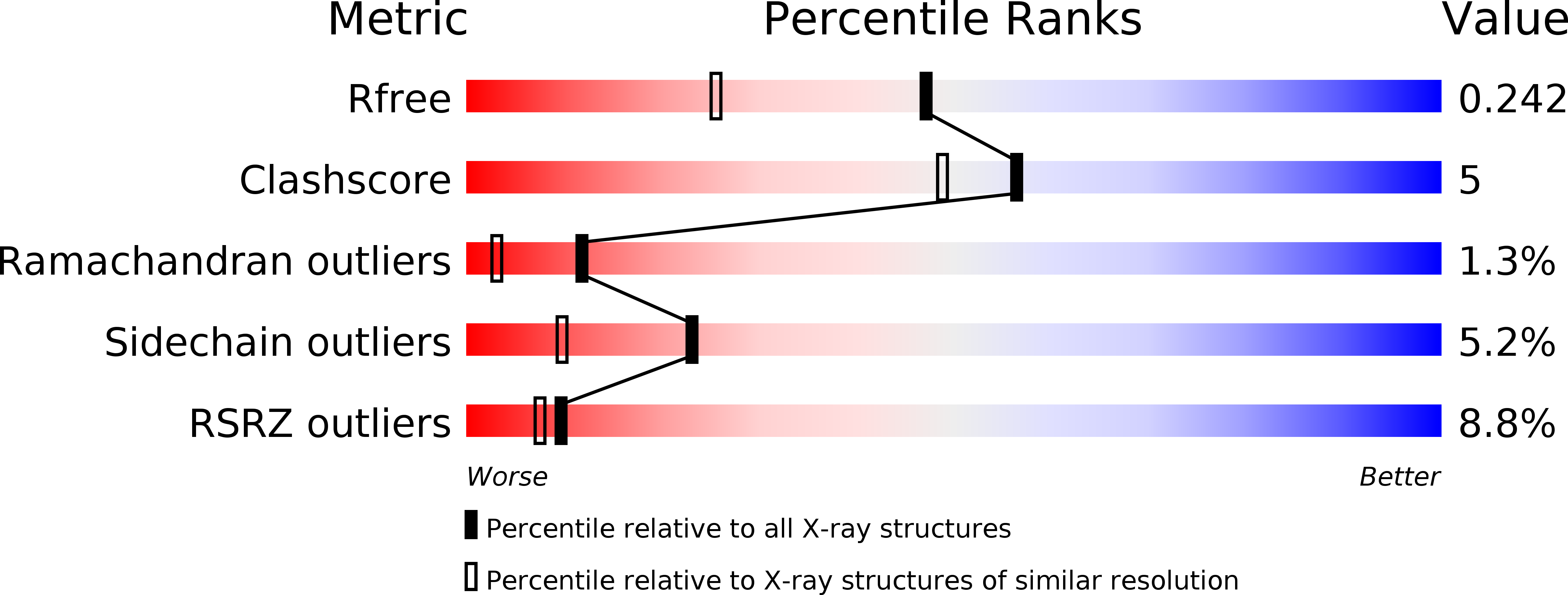
Resolution:
1.80 Å
R-Value Free:
0.24
R-Value Work:
0.20
Space Group:
P 21 21 21
Macromolecular Entities
Structures with similar UniProt ID
Protein Blast
Polymer Type:polypeptide(L)
Molecule:RIBOSOMAL PROTEIN S6 KINASE ALPHA 5
Gene (Uniprot):RPS6KA5
Mutagens:YES
Chain IDs:A
Chain Length:355
Number of Molecules:1
Biological Source:HOMO SAPIENS
Primary Citation
The Structure of Msk1 Reveals a Novel Autoinhibitory Conformation for a Dual Kinase Protein
Structure 12 1066 ? (2004)
PMID: 15274926 DOI: 10.1016/J.STR.2004.02.040

Abstact

Mitogen and stress-activated kinase-1 (MSK1) is a serine/threonine protein kinase that is activated by either p38 or p42ERK MAPKs in response to stress or mitogenic extracellular stimuli. MSK1 belongs to a family of protein kinases that contain two distinct kinase domains in one polypeptide chain. We report the 1.8 A crystal structure of the N-terminal kinase domain of MSK1. The crystal structure reveals a unique inactive conformation with the ATP binding site blocked by the nucleotide binding loop. This inactive conformation is stabilized by the formation of a new three-stranded beta sheet on the N lobe of the kinase domain. The three beta strands come from residues at the N terminus of the kinase domain, what would be the alphaB helix in the active conformation, and the activation loop. The new three-stranded beta sheet occupies a position equivalent to the N terminus of the alphaC helix in active protein kinases.

Legend

Protein

Chemical

Disease

Primary Citation of related structures
Feedback Form
Name
Email
Institute
Feedback