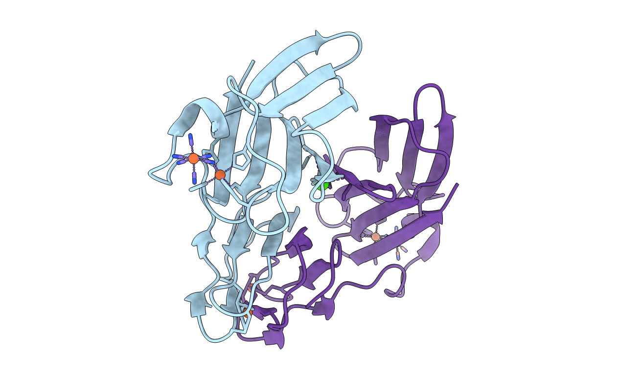
1VZG image
Deposition Date 2004-05-19
Release Date 2004-08-27
Last Version Date 2024-10-16
Entry Detail
PDB ID:
1VZG
Keywords:
Title:
Structure of superoxide reductase bound to ferrocyanide and active site expansion upon X-ray induced photoreduction
Biological Source:
Source Organism:
Host Organism:
Method Details:
Experimental Method:
Resolution:
1.69 Å
R-Value Free:
0.24
R-Value Work:
0.20
R-Value Observed:
0.20
Space Group:
P 21 21 21
Macromolecular Entities
Polymer Type:polypeptide(L)
Molecule:DESULFOFERRODOXIN
Gene (Uniprot):dfx
Mutations:YES
Chain IDs:A, B
Chain Length:126
Number of Molecules:2
Biological Source:DESULFOVIBRIO BAARSII
Primary Citation
Structure of Superoxide Reductase Bound to Ferrocyanide and Active Site Expansion Upon X-Ray Induced Photoreduction
Structure 12 1729 ? (2004)
PMID: 15341736 DOI: 10.1016/J.STR.2004.07.013

Abstact

Some sulfate-reducing and microaerophilic bacteria rely on the enzyme superoxide reductase (SOR) to eliminate the toxic superoxide anion radical (O2*-). SOR catalyses the one-electron reduction of O2*- to hydrogen peroxide at a nonheme ferrous iron center. The structures of Desulfoarculus baarsii SOR (mutant E47A) alone and in complex with ferrocyanide were solved to 1.15 and 1.7 A resolution, respectively. The latter structure, the first ever reported of a complex between ferrocyanide and a protein, reveals that this organo-metallic compound entirely plugs the SOR active site, coordinating the active iron through a bent cyano bridge. The subtle structural differences between the mixed-valence and the fully reduced SOR-ferrocyanide adducts were investigated by taking advantage of the photoelectrons induced by X-rays. The results reveal that photo-reduction from Fe(III) to Fe(II) of the iron center, a very rapid process under a powerful synchrotron beam, induces an expansion of the SOR active site.

Legend

Protein

Chemical

Disease

Primary Citation of related structures