Deposition Date
1992-03-30
Release Date
1994-07-31
Last Version Date
2024-02-14
Entry Detail
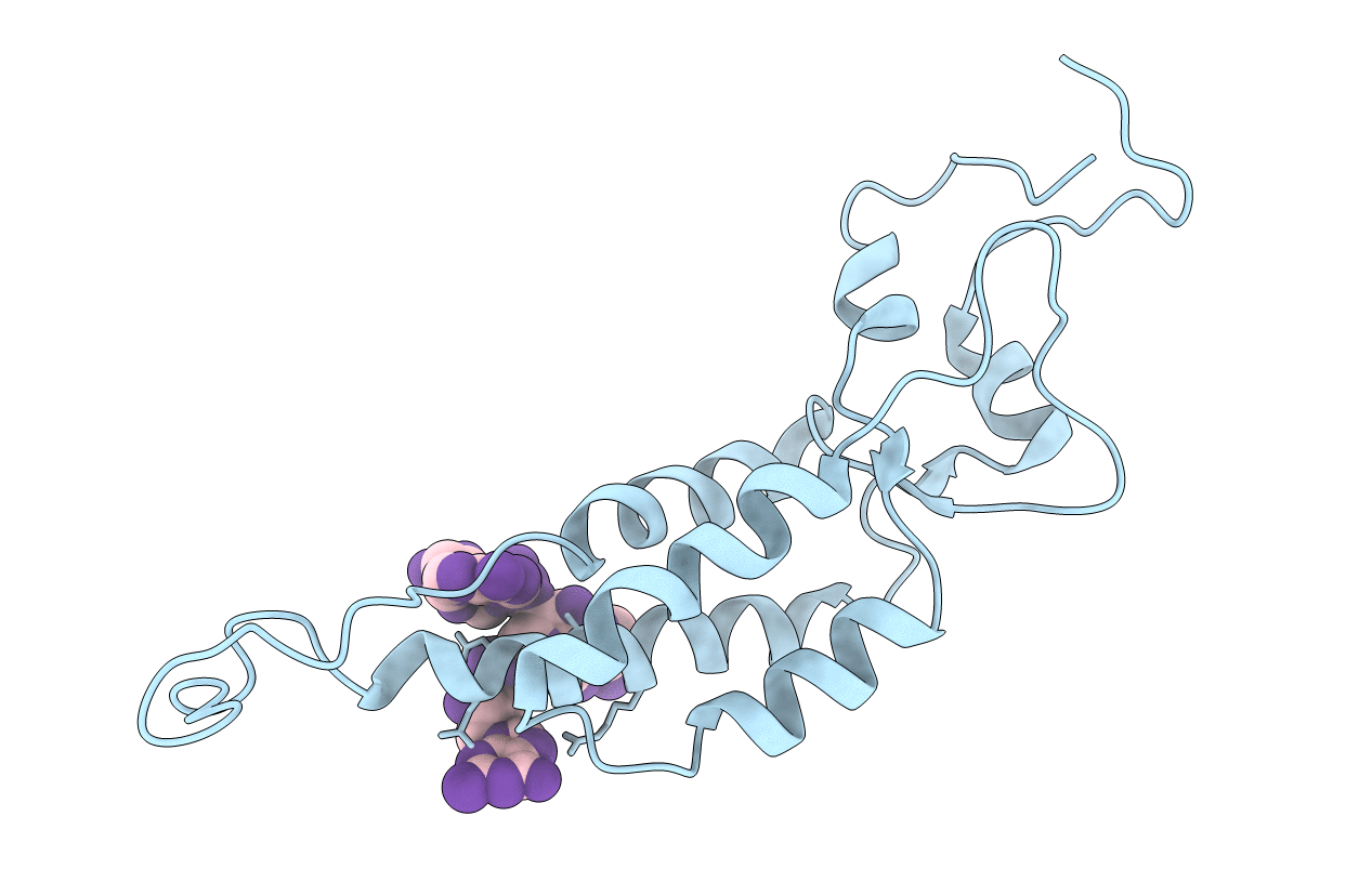
PDB ID:
1VTM
Keywords:
Title:
STRUCTURE OF THE U2 STRAIN OF TOBACCO MOSAIC VIRUS REFINED AT 3.5 ANGSTROMS RESOLUTION USING X-RAY FIBER DIFFRACTION
Biological Source:
Source Organism(s):
Tobacco mild green mosaic virus (TMGMV) (Taxon ID: 12241)
Method Details: