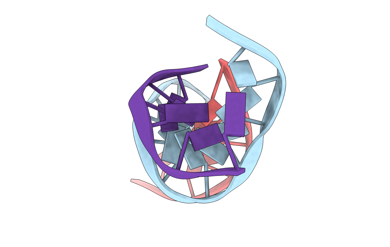
1VTE image
Deposition Date 1990-05-21
Release Date 2011-07-13
Last Version Date 2023-12-27
Entry Detail
PDB ID:
1VTE
Keywords:
Title:
MOLECULAR STRUCTURE OF NICKED DNA. MODEL A4
Biological Source:
Source Organism:
Method Details:
Experimental Method:
Resolution:
3.00 Å
R-Value Observed:
0.18
Space Group:
P 21 21 21
Macromolecular Entities
Polymer Type:polydeoxyribonucleotide
Molecule:DNA (5'-D(*CP*GP*CP*GP*AP*AP*AP*AP*CP*GP*CP*G)-3')
Chain IDs:A
Chain Length:12
Number of Molecules:1
Biological Source:synthetic construct
Polymer Type:polydeoxyribonucleotide
Molecule:DNA (5'-D(*CP*GP*CP*GP*TP*T)-3')
Chain IDs:B
Chain Length:6
Number of Molecules:1
Biological Source:synthetic construct
Polymer Type:polydeoxyribonucleotide
Molecule:DNA (5'-D(*TP*TP*CP*GP*CP*G)-3')
Chain IDs:C
Chain Length:6
Number of Molecules:1
Biological Source:synthetic construct
Primary Citation
Molecular structure of nicked DNA: a substrate for DNA repair enzymes.
Proc. Natl. Acad. Sci. U.S.A. 87 2526 2530 (1990)
PMID: 2320572

Abstact

The molecular structure of a nicked dodecamer DNA double helix, made of a ternary system containing d(CGCGAAAACGCG) + d(CGCGTT) + d(TTCGCG) oligonucleotides, has been determined by x-ray diffraction analysis at 3 A resolution. The molecule adopts a B-DNA conformation, not unlike those found in intact dodecamer DNA molecules crystallized in a somewhat different crystal lattice, despite a gap due to the absence of a phosphate group in the molecule. The helix has a distinct narrow minor groove near the center of the molecule at the AAAA region. This suggests that the internal stabilizing forces due to base stacking and hydrogen-bonding interactions are sufficient to overcome the loss of connectivity associated with the disruption of the covalent backbone of DNA.

Legend

Protein

Chemical

Disease

Primary Citation of related structures