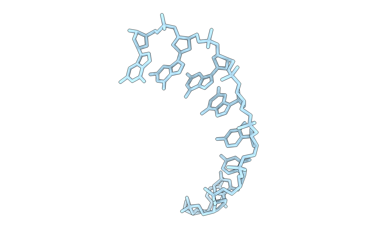
1VT5 image
Deposition Date 1988-08-18
Release Date 2011-07-13
Last Version Date 2023-12-27
Entry Detail
PDB ID:
1VT5
Keywords:
Title:
THE CRYSTAL STRUCTURE OF D(CCCCGGGG): A NEW A-FORM VARIANT WITH AN EXTENDED BACKBONE CONFORMATION
Biological Source:
Source Organism:
(Taxon ID: )
Method Details:
Experimental Method:
Resolution:
2.25 Å
R-Value Work:
0.24
R-Value Observed:
0.24
Space Group:
P 43 21 2
Macromolecular Entities
Polymer Type:polydeoxyribonucleotide
Molecule:DNA (5'-D(*CP*CP*CP*CP*GP*GP*GP*G)-3')
Chain IDs:A
Chain Length:8
Number of Molecules:1
Biological Source:
Ligand Molecules
Primary Citation
The crystal structure of d(CCCCGGGG): a new A-form variant with an extended backbone conformation.
J.Biomol.Struct.Dyn. 5 199 217 (1987)
PMID: 3271472

Abstact

The crystal structure of d(CCCCGGGG) has been determined at a resolution of 2.25 A. The oligomers crystallize as A-DNA duplexes occupying crystallographic two-fold axes. The backbone conformation is, in general, similar to that observed in previously reported crystal structures of A-DNA fragments, except for the central linkage, where it adopts an extended structure resulting from all trans conformation at the P-O5'-C5'-C4' bonds. This type of conformation facilitates interstrand stacking between the guanines at the C-G site. The local helix twist at this step is very small (25 degrees) compared to an overall average of 33.5 degrees. The unique structure of the C-G base-pair step, namely the extended backbone and the distinct stacking geometry, may be an important feature in the recognition mechanism between double-stranded DNA molecules and restriction endonucleases such as Msp I, which cuts the sequence CCGG very specifically with a rate unaffected by neighboring base pairs.

Legend

Protein

Chemical

Disease

Primary Citation of related structures