1VS0 image
Deposition Date 2006-01-27
Release Date 2006-02-28
Last Version Date 2025-03-26
Entry Detail
PDB ID:
1VS0
Keywords:
Title:
Crystal Structure of the Ligase Domain from M. tuberculosis LigD at 2.4A
Biological Source:
Source Organism:
Host Organism:
Method Details:
Experimental Method:
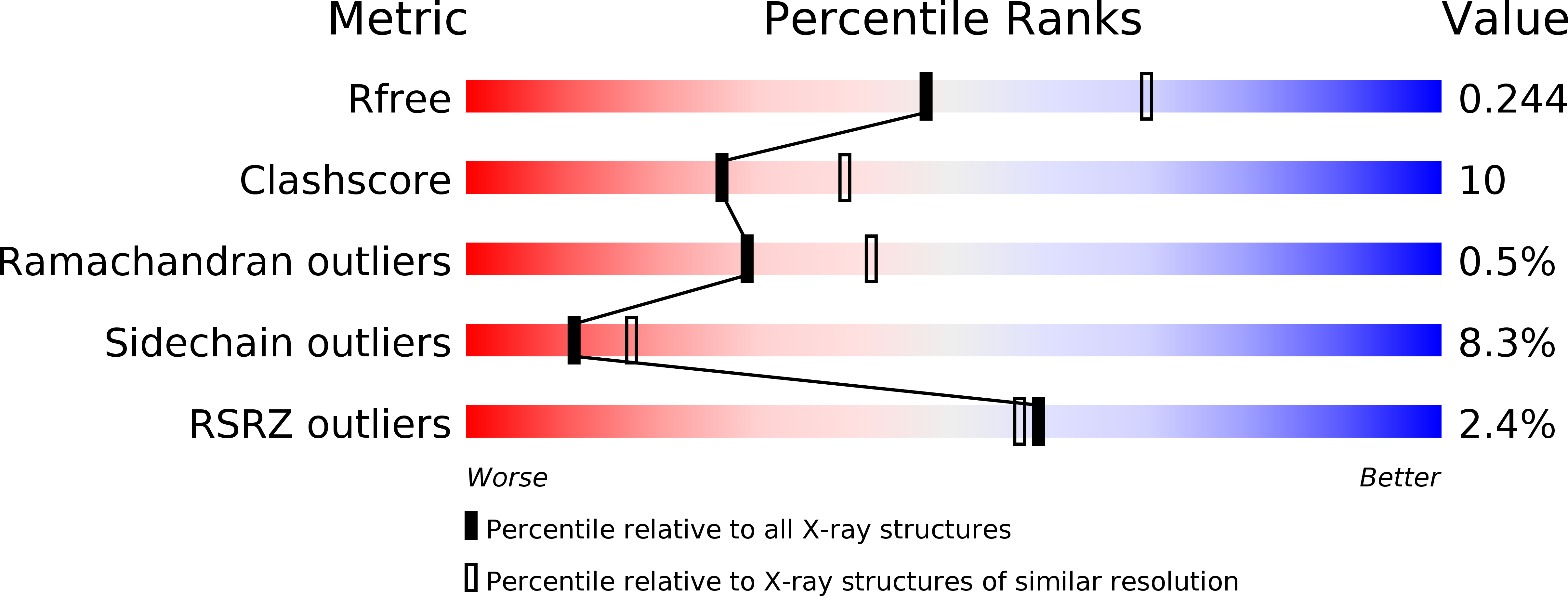
Resolution:
2.40 Å
R-Value Free:
0.24
R-Value Work:
0.19
R-Value Observed:
0.19
Space Group:
P 32 2 1
Macromolecular Entities
Polymer Type:polypeptide(L)
Molecule:Putative DNA ligase-like protein Rv0938/MT0965
Chain IDs:A, B
Chain Length:310
Number of Molecules:2
Biological Source:Mycobacterium tuberculosis
Modified Residue
Compound ID Chain ID Parent Comp ID Details 2D Image
APK A LYS ?
MSE A MET SELENOMETHIONINE
Primary Citation
Crystal Structure and Nonhomologous End-joining Function of the Ligase Component of Mycobacterium DNA Ligase D.
J.Biol.Chem. 281 13412 13423 (2006)
PMID: 16476729 DOI: 10.1074/jbc.M513550200

Abstact

DNA ligase D (LigD) is a large polyfunctional enzyme involved in nonhomologous end-joining (NHEJ) in mycobacteria. LigD consists of a C-terminal ATP-dependent ligase domain fused to upstream polymerase and phosphoesterase modules. Here we report the 2.4 angstroms crystal structure of the ligase domain of Mycobacterium LigD, captured as the covalent ligase-AMP intermediate with a divalent metal in the active site. A chloride anion on the protein surface coordinated by the ribose 3'-OH and caged by arginine and lysine side chains is a putative mimetic of the 5'-phosphate at a DNA nick. Structure-guided mutational analysis revealed distinct requirements for the adenylylation and end-sealing reactions catalyzed by LigD. We found that a mutation of Mycobacterium LigD that ablates only ligase activity results in decreased fidelity of NHEJ in vivo and a strong bias of mutagenic events toward deletions instead of insertions at the sealed DNA ends. This phenotype contrasts with the increased fidelity of double-strand break repair in deltaligD cells or in a strain in which only the polymerase function of LigD is defective. We surmise that the signature error-prone quality of bacterial NHEJ in vivo arises from a dynamic balance between the end-remodeling and end-sealing steps.

Legend

Protein

Chemical

Disease

Primary Citation of related structures