1VRO image
Deposition Date 2005-04-14
Release Date 2005-04-19
Last Version Date 2023-12-27
Entry Detail
PDB ID:
1VRO
Keywords:
Title:
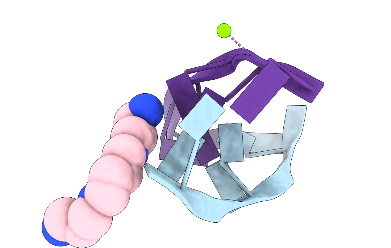
Selenium-Assisted Nucleic Acid Crystallography: Use of Phosphoroselenoates for MAD Phasing of a DNA Structure
Biological Source:
Source Organism:
(Taxon ID: )
Method Details:
Experimental Method:
Resolution:
1.10 Å
R-Value Free:
0.12
R-Value Work:
0.09
Space Group:
P 21 21 21
Macromolecular Entities
Polymer Type:polydeoxyribonucleotide
Molecule:5'-D(*CP*(GMS)P*CP*GP*CP*G)-3'
Chain IDs:A, B
Chain Length:6
Number of Molecules:2
Biological Source:
Modified Residue
Compound ID Chain ID Parent Comp ID Details 2D Image
GMS A DG ?
Primary Citation
Selenium-Assisted Nucleic Acid Crystallography: Use of Phosphoroselenoates for MAD Phasing of a DNA Structure
J.Am.Chem.Soc. 124 14910 14916 (2002)
PMID: 12475332 DOI: 10.1021/ja021058b

Abstact

The combination of synchrotron radiation and a variety of atoms or ions (either covalently attached to the biomolecule prior to crystallization or soaked into crystals) that serve as anomalous scatterers constitutes a powerful tool in the X-ray crystallographer's repertoire of structure determination techniques. Phosphoroselenoates in which one of the nonbridging phosphate oxygens in the backbone is replaced by selenium offer a simplified means for introducing an anomalous scatterer into oligonucleotides by conventional solid-phase synthesis. Unlike other methods that are used to derivatize DNA or RNA by covalent attachment of a heavy atom (i.e., bromine at the C5 position of pyrimidines), tedious synthesis of specialized nucleosides is not required. Introduction of selenium is readily accomplished in solid-phase oligonucleotide synthesis by replacing the standard oxidation agent with a solution of potassium selenocyanide. This results in a diastereomeric mixture of phosphoroselenoates that can be separated by strong anion-exchange HPLC. As a test case, all 10 DNA hexamers of the sequence CGCGCG containing a single phosphoroselenoate linkage (PSe) were prepared. Crystals were grown for a subset of them, and the structure of [d(C(PSe)GCGCG)](2) was determined by the multiwavelength anomalous dispersion technique and refined to 1.1 A resolution.

Legend

Protein

Chemical

Disease

Primary Citation of related structures