
Deposition Date
2005-02-23
Release Date
2005-03-22
Last Version Date
2024-11-20
Entry Detail
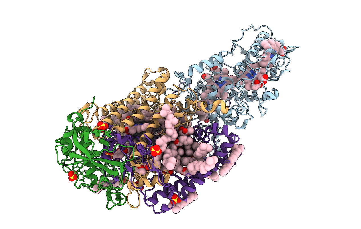
PDB ID:
1VRN
Keywords:
Title:
PHOTOSYNTHETIC REACTION CENTER BLASTOCHLORIS VIRIDIS (ATCC)
Biological Source:
Source Organism(s):
Blastochloris viridis (Taxon ID: 1079)
Method Details:
Experimental Method:
Resolution:
2.20 Å
R-Value Free:
0.21
R-Value Work:
0.19
R-Value Observed:
0.19
Space Group:
P 43 21 2
