1VRK image
Deposition Date 1997-09-24
Release Date 1999-04-27
Last Version Date 2024-10-30
Entry Detail
PDB ID:
1VRK
Title:
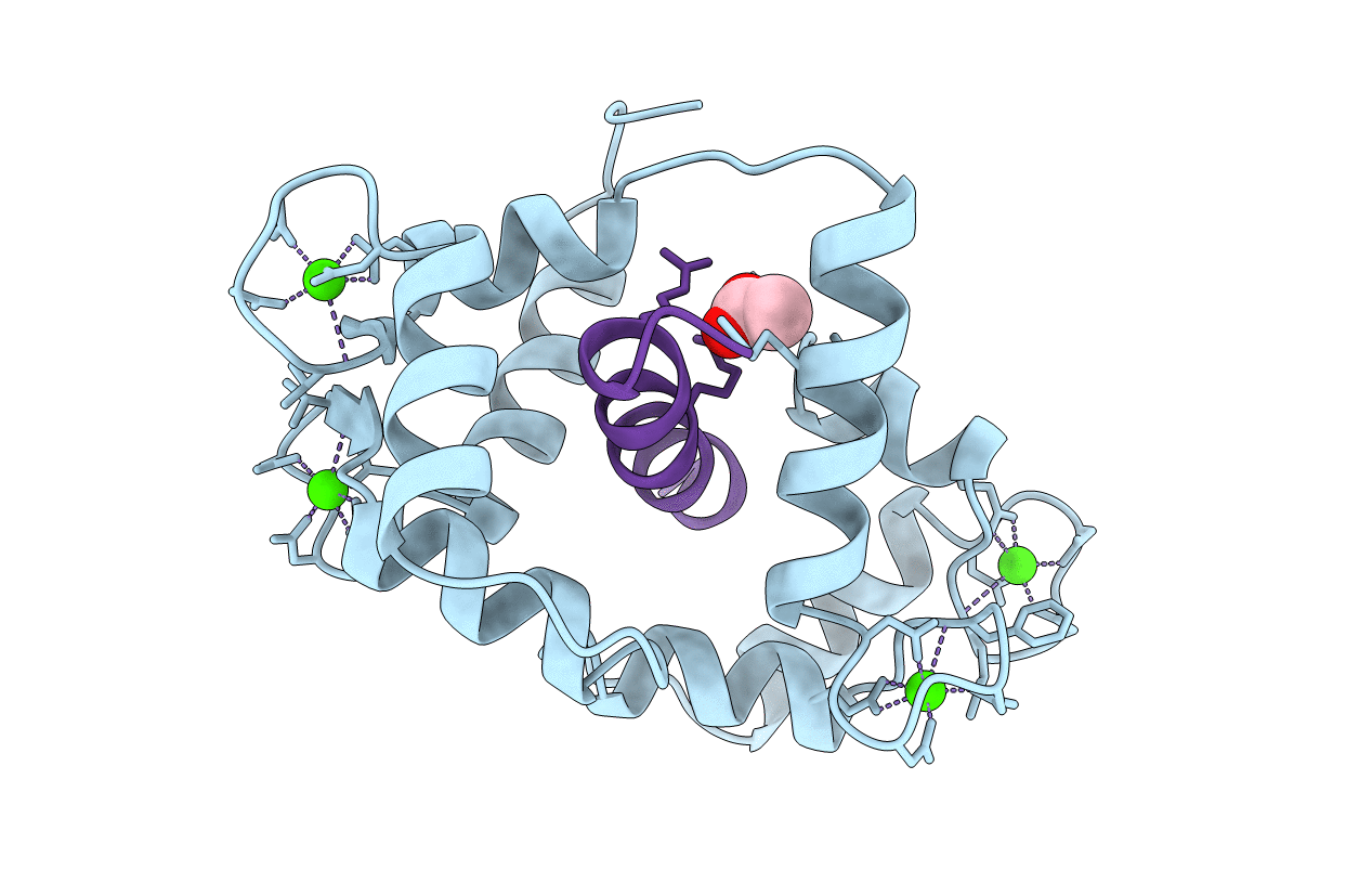
THE 1.9 ANGSTROM STRUCTURE OF E84K-CALMODULIN RS20 PEPTIDE COMPLEX
Biological Source:
Source Organism:
Host Organism:
Method Details:
Experimental Method:
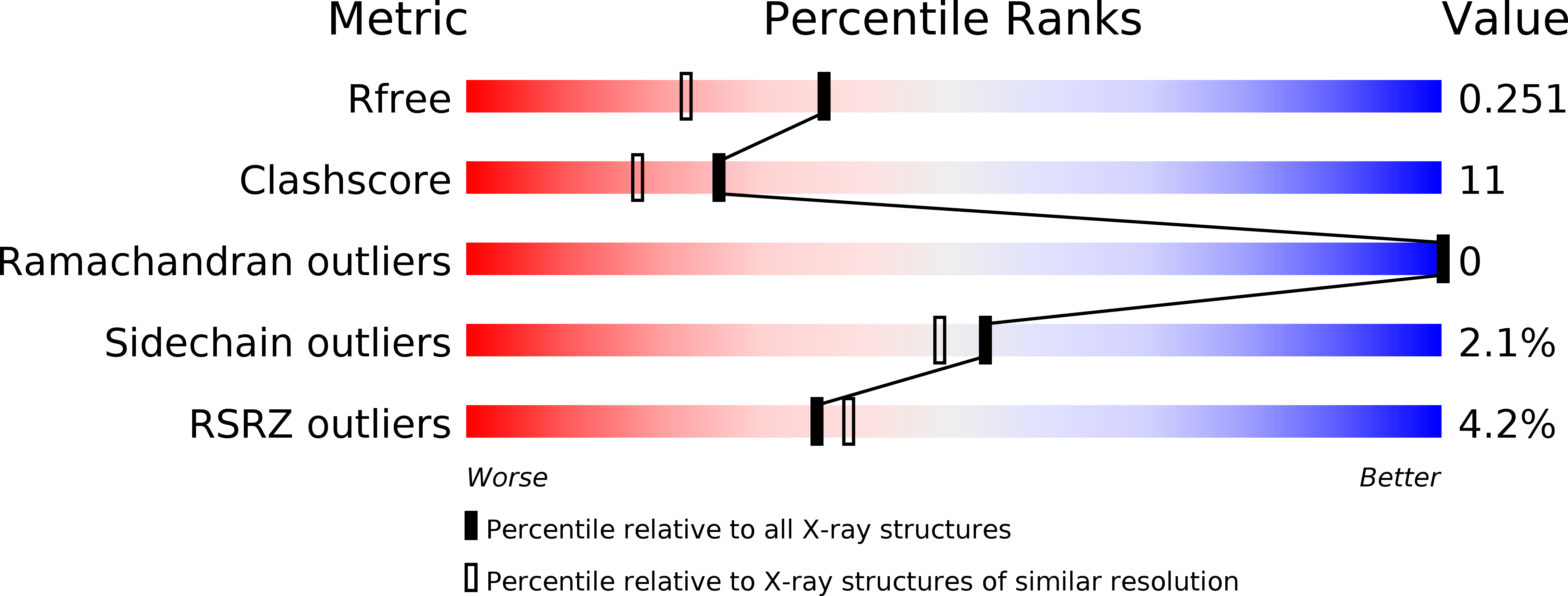
Resolution:
1.90 Å
R-Value Free:
0.24
R-Value Work:
0.17
R-Value Observed:
0.17
Space Group:
P 1 21 1
Macromolecular Entities
Polymer Type:polypeptide(L)
Molecule:CALMODULIN
Mutagens:E84K
Chain IDs:A
Chain Length:148
Number of Molecules:1
Biological Source:synthetic construct
Modified Residue
Compound ID Chain ID Parent Comp ID Details 2D Image
TRF B TRP N1-FORMYL-TRYPTOPHAN
Primary Citation
Analysis of the functional coupling between calmodulin's calcium binding and peptide recognition properties.
Biochemistry 38 3936 3947 (1999)
PMID: 10194305 DOI: 10.1021/bi9821263

Abstact

The enhancement of calmodulin's (CaM) calcium binding activity by an enzyme or a recognition site peptide and its diminution by key point mutations at the protein recognition interface (e.g., E84K-CaM), which is more than 20 A away from the nearest calcium ligation structure, can be described by an expanded version of the Adair-Klotz equation for multiligand binding. The expanded equation can accurately describe the calcium binding events and their variable linkage to protein recognition events can be extended to other CaM-regulated enzymes and can potentially be applied to a diverse array of ligand binding systems with allosteric regulation of ligand binding, whether by other ligands or protein interaction. The 1.9 A resolution X-ray crystallographic structure of the complex between E84K-CaM and RS20 peptide, the CaM recognition site peptide from vertebrate smooth muscle and nonmuscle forms of myosin light chain kinase, provides insight into the structural basis of the functional communication between CaM's calcium ligation structures and protein recognition surfaces. The structure reveals that the complex adapts to the effect of the functional mutation by discrete adjustments in the helix that contains E84. This helix is on the amino-terminal side of the helix-loop-helix structural motif that is the first to be occupied in CaM's calcium binding mechanism. The results reported here are consistent with a sequential and cooperative model of CaM's calcium binding activity in which the two globular and flexible central helix domains are functionally linked, and provide insight into how CaM's calcium binding activity and peptide recognition properties are functionally coupled.

Legend

Protein

Chemical

Disease

Primary Citation of related structures
Feedback Form
Name
Email
Institute
Feedback