
Deposition Date
2005-02-21
Release Date
2005-04-19
Last Version Date
2023-12-27
Entry Detail
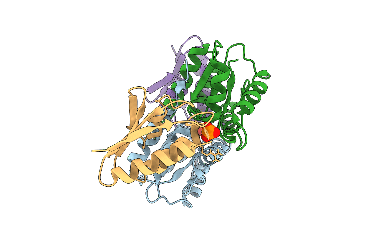
PDB ID:
1VRC
Keywords:
Title:
Complex of enzyme IIAmannose and the histidine-containing phosphocarrier protein HPr from escherichia coli nmr, restrained regularized mean structure
Biological Source:
Source Organism(s):
Escherichia coli (Taxon ID: 562)
Expression System(s):
Method Details:
Experimental Method:
Conformers Calculated:
100
Conformers Submitted:
2
Selection Criteria:
REGULARIZED MEAN STRUCTURES
