1VQX image
Deposition Date 2005-01-07
Release Date 2005-01-18
Last Version Date 2024-10-09
Entry Detail
PDB ID:
1VQX
Title:
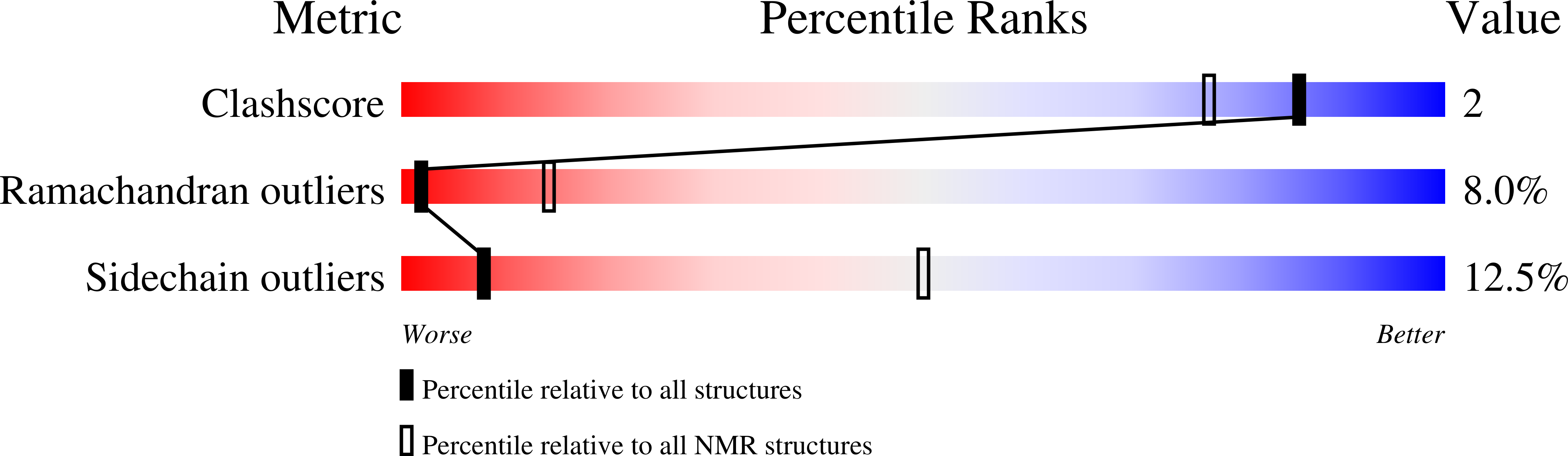
ARRESTIN-BOUND NMR STRUCTURES OF THE PHOSPHORYLATED CARBOXY-TERMINAL DOMAIN OF RHODOPSIN, REFINED
Biological Source:
Source Organism:
(Taxon ID: )
Method Details:
Experimental Method:
Conformers Calculated:
100
Conformers Submitted:
15
Selection Criteria:
structures with the lowest energy
Macromolecular Entities
Polymer Type:polypeptide(L)
Molecule:RHODOPSIN
Gene (Uniprot):RHO
Chain IDs:A
Chain Length:19
Number of Molecules:1
Biological Source:
Modified Residue
Compound ID Chain ID Parent Comp ID Details 2D Image
SEP A SER PHOSPHOSERINE
TPO A THR PHOSPHOTHREONINE
Ligand Molecules
Primary Citation
Conformational Changes in the Phosphorylated C-Terminal Domain of Rhodopsin During Rhodopsin Arrestin Interactions
J.Biol.Chem. 279 51203 ? (2004)
PMID: 15351781 DOI: 10.1074/jbc.M407341200

Abstact

Phosphorylation of activated G-protein-coupled receptors and the subsequent binding of arrestin mark major molecular events of homologous desensitization. In the visual system, interactions between arrestin and the phosphorylated rhodopsin are pivotal for proper termination of visual signals. By using high resolution proton nuclear magnetic resonance spectroscopy of the phosphorylated C terminus of rhodopsin, represented by a synthetic 7-phosphopolypeptide, we show that the arrestin-bound conformation is a well ordered helix-loop structure connected to rhodopsin via a flexible linker. In a model of the rhodopsin-arrestin complex, the phosphates point in the direction of arrestin and form a continuous negatively charged surface, which is stabilized by a number of positively charged lysine and arginine residues of arrestin. Opposite to the mostly extended structure of the unphosphorylated C-terminal domain of rhodopsin, the arrestin-bound C-terminal helix is a compact domain that occupies a central position between the cytoplasmic loops and occludes the key binding sites of transducin. In conjunction with other binding sites, the helix-loop structure provides a mechanism of shielding phosphates in the center of the rhodopsin-arrestin complex and appears critical in guiding arrestin for high affinity binding with rhodopsin.

Legend

Protein

Chemical

Disease

Primary Citation of related structures