1VPR image
Deposition Date 2004-11-15
Release Date 2005-02-08
Last Version Date 2024-10-30
Entry Detail
PDB ID:
1VPR
Title:
Crystal structure of a luciferase domain from the dinoflagellate Lingulodinium polyedrum
Biological Source:
Source Organism:
Host Organism:
Method Details:
Experimental Method:
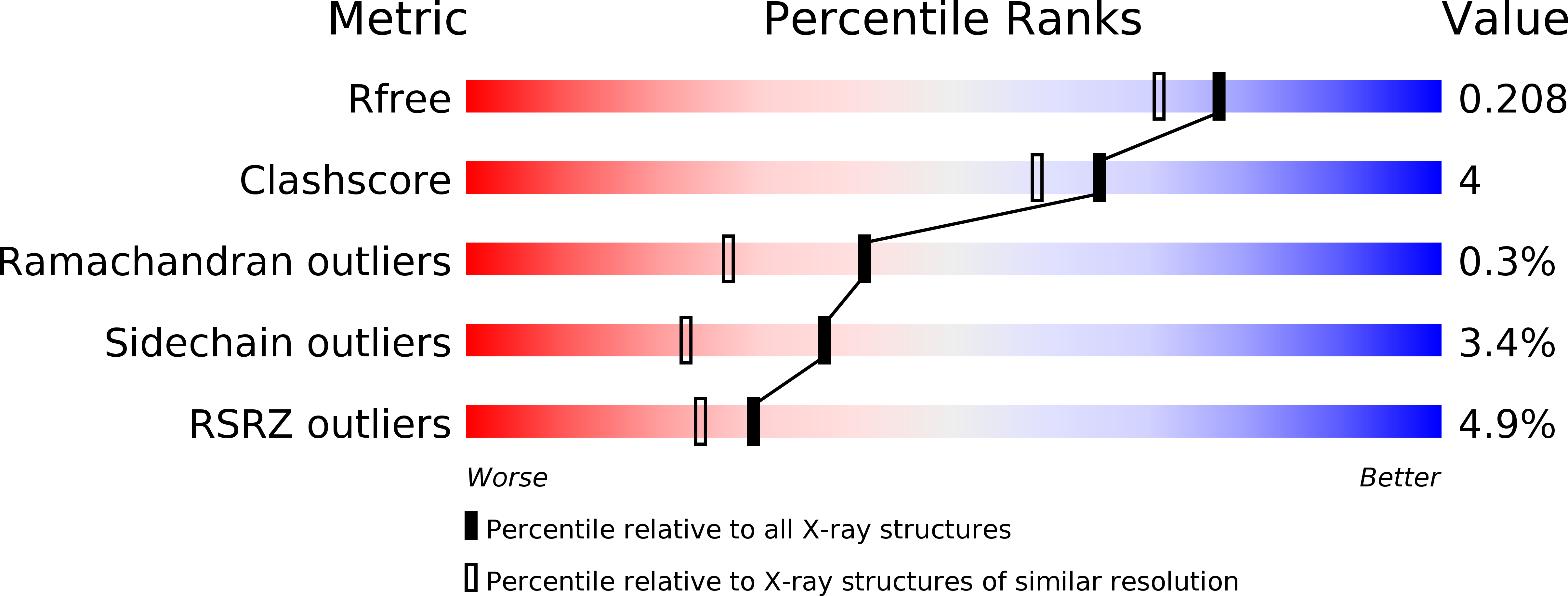
Resolution:
1.80 Å
R-Value Free:
0.23
R-Value Work:
0.19
R-Value Observed:
0.19
Space Group:
P 21 21 21
Macromolecular Entities
Polymer Type:polypeptide(L)
Molecule:luciferase
Chain IDs:A
Chain Length:374
Number of Molecules:1
Biological Source:Lingulodinium polyedrum
Modified Residue
Compound ID Chain ID Parent Comp ID Details 2D Image
MSE A MET SELENOMETHIONINE
Primary Citation
Crystal structure of a pH-regulated luciferase catalyzing the bioluminescent oxidation of an open tetrapyrrole
Proc.Natl.Acad.Sci.USA 102 1378 1383 (2005)
PMID: 15665092 DOI: 10.1073/pnas.0409335102

Abstact

The luciferase of Lingulodinium polyedrum, a marine bioluminescent dinoflagellate, consists of three similar but not identical domains in a single polypeptide. Each encodes an active luciferase that catalyzes the oxidation of a chlorophyll-derived open tetrapyrrole (dinoflagellate luciferin) to produce blue light. These domains share no sequence similarity with any other in the GenBank database and no structural or motif similarity with any other luciferase. We report here the 1.8-A crystal structure of the third domain, D3, at pH 8, and a mechanism for its activity regulation by pH. D3 consists of two major structural elements: a beta-barrel pocket putatively for substrate binding and catalysis and a regulatory three-helix bundle. N-terminal histidine residues previously shown to regulate activity by pH are at the interface of the helices in the bundle. Molecular dynamics calculations indicate that, in response to changes in pH, these histidines could trigger a large molecular motion of the bundle, thereby exposing the active site to the substrate.

Legend

Protein

Chemical

Disease

Primary Citation of related structures