1VNA image
Deposition Date 1993-10-26
Release Date 1994-01-31
Last Version Date 2024-10-09
Entry Detail
PDB ID:
1VNA
Keywords:
Title:
PROTON NUCLEAR MAGNETIC RESONANCE AND DISTANCE GEOMETRY(SLASH)SIMULATED ANNEALING STUDIES ON THE VARIANT-1 NEUROTOXIN FROM THE NEW WORLD SCORPION CENTRUROIDES SCULPTURATUS EWING
Biological Source:
Source Organism:
Method Details:
Experimental Method:
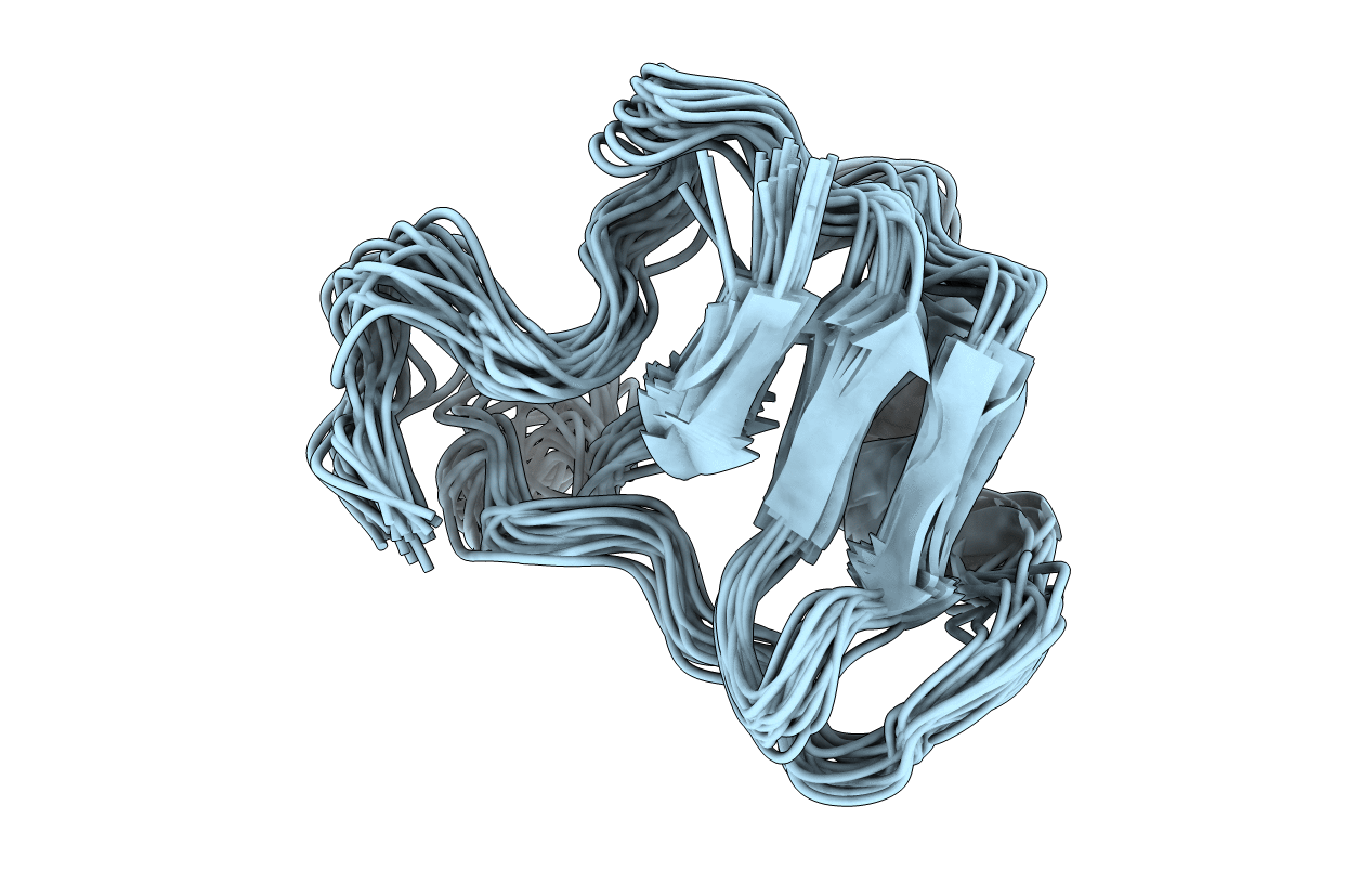
Conformers Submitted:
26
Macromolecular Entities
Polymer Type:polypeptide(L)
Molecule:NEUROTOXIN
Chain IDs:A
Chain Length:65
Number of Molecules:1
Biological Source:Centruroides sculpturatus
Ligand Molecules
Primary Citation
Proton nuclear magnetic resonance and distance geometry/simulated annealing studies on the variant-1 neurotoxin from the New World scorpion Centruroides sculpturatus Ewing.
Biochemistry 33 2468 2475 (1994)
PMID: 8117707 DOI: 10.1021/bi00175a015

Abstact

The sequence-specific proton resonance assignments for the variant-1 (CsE-v1) neurotoxin from the venom of the New World scorpion Centruroides sculpturatus Ewing (range Southwestern United States) have been performed by 2D 1H NMR spectroscopy at 600 MHz. The stereospecific assignments for the beta-methylene protons of 19 non-proline residues have been determined. A number of short-, medium-, and long-range NOESY contacts as well as the backbone and the side-chain vicinal coupling constants for several residues have been determined. Slowly exchanging amide hydrogens from a number of residues have been identified. On the basis of the NMR data, the solution structure of this protein has been determined by a hybrid procedure consisting of distance geometry and dynamical simulated annealing refinement calculations. Distance constraints from the NOESY data and torsion angle constraints from proton vicinal coupling constant data were used in the simulated annealing calculations. The three-dimensional structure of CsE-v1 is characterized by a three-stranded antiparallel beta-sheet, a short alpha-helix, a cis-proline, and intervening loops. A comparison with the solution NMR data of a homologous protein (CsE-v3) from the Centruroides venom, shows that the structures are essentially similar, except for some minor differences. Some of the NMR spectral perturbations are felt in regions far removed from sites of amino acid substitutions. The hydrophobic surface in CsE-v1 is slightly more extended than in CsE-v3.

Legend

Protein

Chemical

Disease

Primary Citation of related structures