Abstact
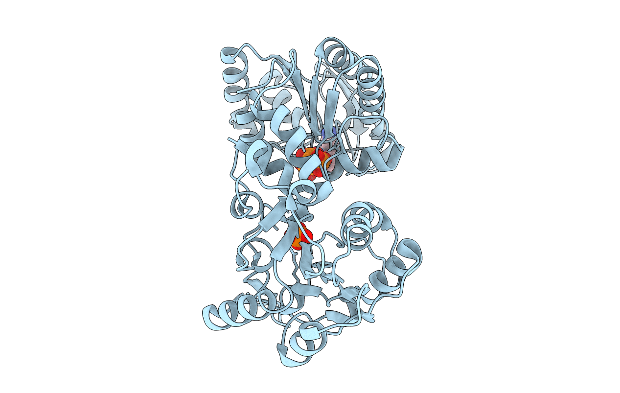
The complexes of pig muscle 3-phosphoglycerate kinase with the substrate MgATP and with the nonsubstrate Mg(2+)-free ATP have been characterized by binding, kinetic, and crystallographic studies. Comparative experiments with ADP and MgADP have also been carried out. In contrast to the less specific and largely ionic binding of Mg(2+)-free ATP and ADP, specific occupation of the adenosine binding pocket by MgATP and MgADP has been revealed by displacement experiments with adenosine and anions, as well as supported by isothermal calorimetric titrations. The Mg(2+)-free nucleotides similarly stabilize the overall protein structure and restrict the conformational flexibility around the reactive thiol groups of helix 13, as observed by differential scanning microcalorimetry and thiol reactivity studies, respectively. The metal complexes, however, behave differently. MgADP, but not MgATP, further increases the conformational stability with respect to its Mg(2+)-free form, which indicates their different modes of binding to the enzyme. Crystal structures of the binary complexes of the enzyme with MgATP and with ATP (2.1 and 1.9 A resolution, respectively) have shown that the orientation and interaction of phosphates of MgATP largely differ not only from those of ATP but also from the previously determined ones of either MgADP [Davies, G. J., Gamblin, S. J., Littlechild, J. A., Dauter, Z., Wilson, K. S., and Watson, H. C. (1994) Acta Crystallogr. D50, 202-209] or the metal complexes of AMP-PNP [May, A., Vas, M., Harlos, K., and Blake, C. C. F. (1996) Proteins 24, 292-303; Auerbach, G., Huber, R., Grattinger, M., Zaiss, K., Schurig, H., Jaenicke, R., and Jacob, U. (1997) Structure 5, 1475-1483] and are more similar to the interactions formed with MgAMP-PCP [Kovári, Z., Flachner, B., Náray-Szabó, G., and Vas, M. (2002) Biochemistry 41, 8796-8806]. Mg(2+) is liganded to both beta- and gamma-phosphates of ATP, while beta-phosphate is linked to the conserved Asp218, i.e., to the N-terminus of helix 8, through a water molecule; the known interactions of either MgADP or the metal complexes of AMP-PNP with the N-terminus of helix 13 and with Asn336 of beta-strand J are absent in the case of MgATP. Fluctuation of MgATP phosphates between two alternative sites has been proposed to facilitate the correct positioning of the mobile side chain of Lys215, and the catalytically competent active site is thereby completed.