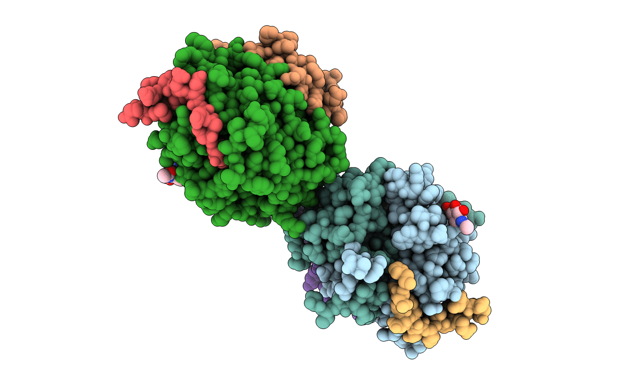
1VIT image
Deposition Date 1996-01-31
Release Date 1997-04-21
Last Version Date 2024-11-13
Entry Detail
PDB ID:
1VIT
Title:
THROMBIN:HIRUDIN 51-65 COMPLEX
Biological Source:
Source Organism:
Bos taurus (Taxon ID: 9913)
Method Details:
Experimental Method:
Resolution:
3.20 Å
R-Value Work:
0.19
R-Value Observed:
0.19
Space Group:
P 61 2 2
Macromolecular Entities
Polymer Type:polypeptide(L)
Molecule:EPSILON THROMBIN
Gene (Uniprot):F2
Chain IDs:E (auth: F)
Chain Length:150
Number of Molecules:1
Biological Source:Bos taurus
Polymer Type:polypeptide(L)
Molecule:EPSILON THROMBIN
Gene (Uniprot):F2
Chain IDs:F (auth: G)
Chain Length:109
Number of Molecules:1
Biological Source:Bos taurus
Polymer Type:polypeptide(L)
Molecule:ALPHA THROMBIN
Gene (Uniprot):F2
Chain IDs:B (auth: H)
Chain Length:259
Number of Molecules:1
Biological Source:Bos taurus
Polymer Type:polypeptide(L)
Molecule:EPSILON THROMBIN
Gene (Uniprot):F2
Chain IDs:A (auth: L), D (auth: M)
Chain Length:49
Number of Molecules:2
Biological Source:Bos taurus
Modified Residue
Compound ID Chain ID Parent Comp ID Details 2D Image
ASN B ASN GLYCOSYLATION SITE
Ligand Molecules
Primary Citation
Structure of a bovine thrombin-hirudin51-65 complex determined by a combination of molecular replacement and graphics. Incorporation of known structural information in molecular replacement.
Acta Crystallogr.,Sect.D 52 453 464 (1996)
PMID: 15299666 DOI: 10.1107/S0907444996000364

Abstact

Crystals of the bovine thrombin-hirudins(51-65) complex have space group P6(1)22 with cell constants a = 116.4, and c = 200.6 A and two thrombin molecules in the asymmetric unit. Only one thrombin molecule could be located by generalized molecular replacement; the second was fit visually as a rigid body to an improved electron-density difference map. The structure was refined to R = 0.192 with two B values per residue (main chain and side chain) at 3.2 A. The polar interactions of the peptides with the exosite of thrombin show differences consistent with the known flexibility in the interactions of the C-terminal peptide of hirudin with thrombin. The hirudin peptide in complex 2 has a higher temperature factor as compared with peptide 1 which may be correlated partly with a larger number of short-range electrostatic interactions between peptide 1 and thrombin and partly with the fact that thrombin 2 is epsilon-thrombin which is cleaved at Thr149A near the peptide binding site. Later, using this structure as a test case, it was shown that the position for the second thrombin could also be determined by a novel modification of the molecular-replacement method in which the contribution of the known molecule is subtracted from the structure factors. This approach is facile and applicable to any crystal containing two or more macromolecules in the asymmetric unit in which some but not all of the molecules can be determined by molecular replacement.

Legend

Protein

Chemical

Disease

Primary Citation of related structures