Deposition Date
1999-04-26
Release Date
1999-05-06
Last Version Date
2023-12-27
Entry Detail
PDB ID:
1VFY
Keywords:
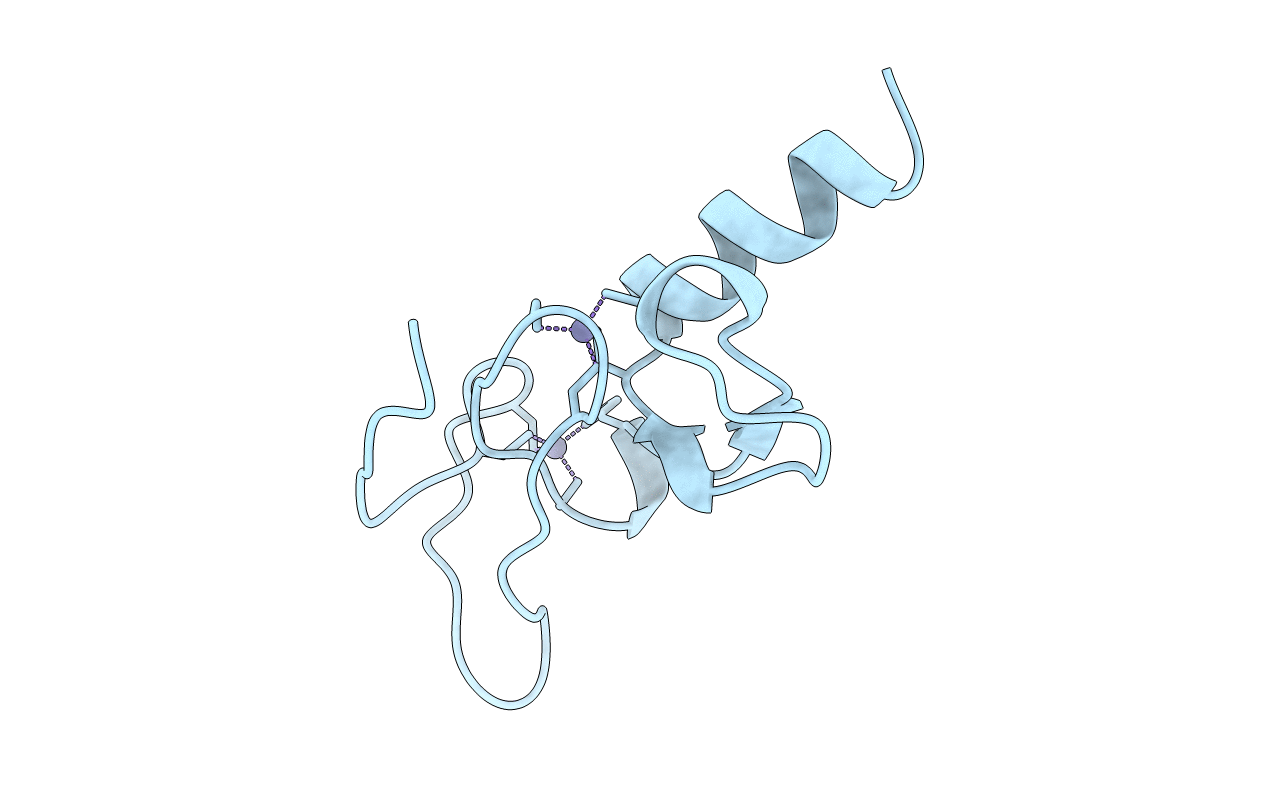
Title:
PHOSPHATIDYLINOSITOL-3-PHOSPHATE BINDING FYVE DOMAIN OF VPS27P PROTEIN FROM SACCHAROMYCES CEREVISIAE
Biological Source:
Source Organism(s):
Saccharomyces cerevisiae (Taxon ID: 4932)
Expression System(s):
Method Details:
Experimental Method:
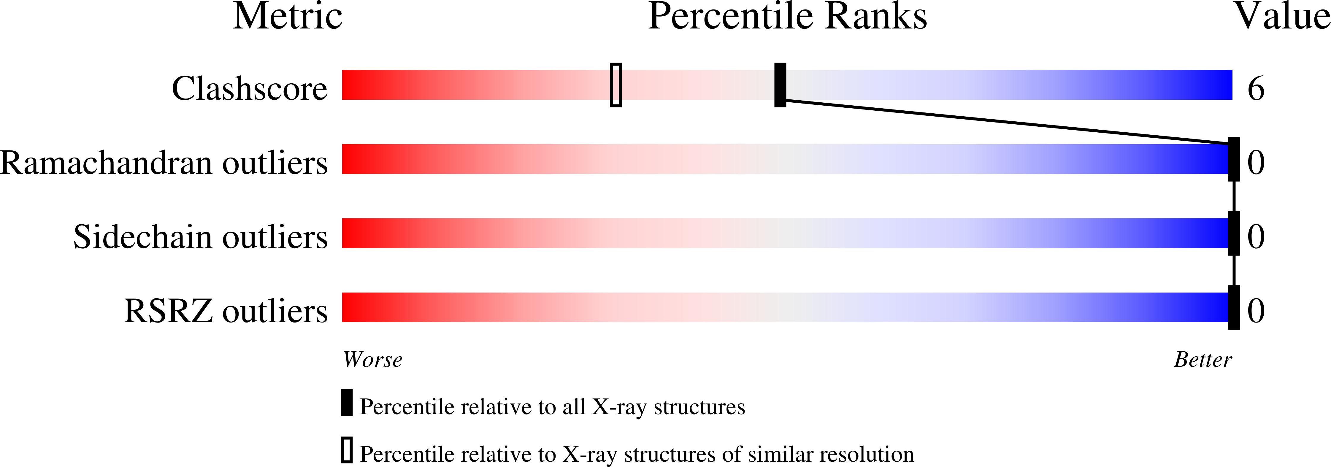
Resolution:
1.15 Å
R-Value Free:
0.18
R-Value Work:
0.17
Space Group:
P 1