1VFW image
Deposition Date 2004-04-19
Release Date 2004-08-10
Last Version Date 2023-12-27
Entry Detail
PDB ID:
1VFW
Title:
Crystal Structure of the Kif1A Motor Domain Complexed With Mg-AMPPNP
Biological Source:
Source Organism:
Mus musculus (Taxon ID: 10090)
Host Organism:
Method Details:
Experimental Method:
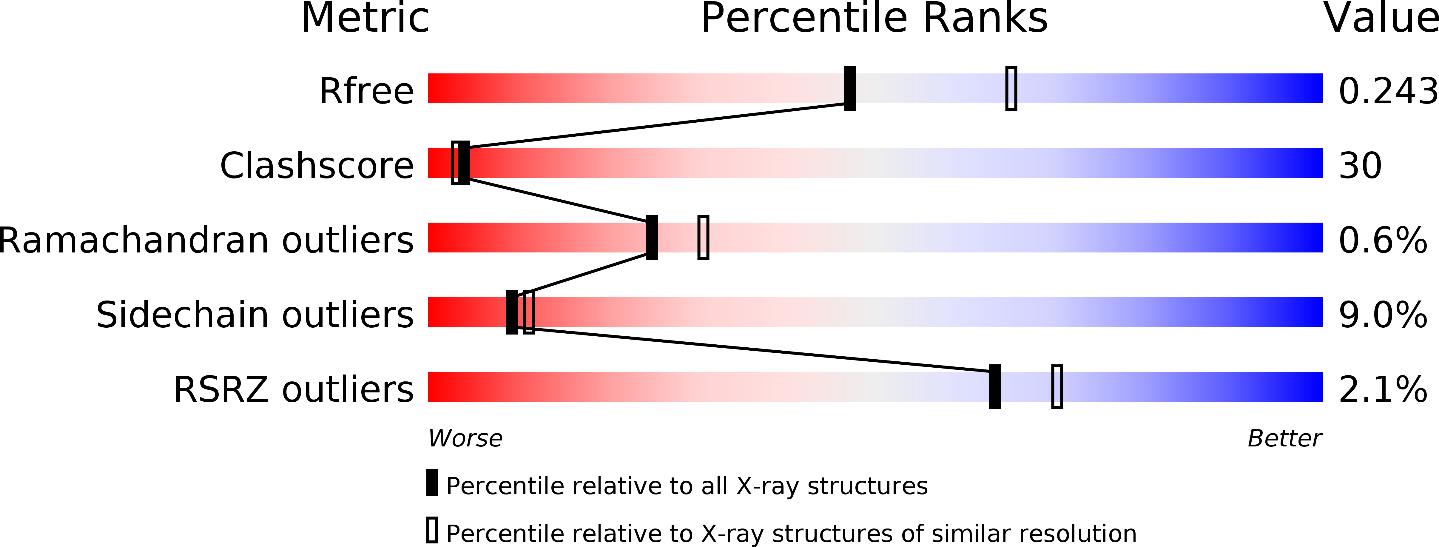
Resolution:
2.30 Å
R-Value Free:
0.24
R-Value Work:
0.21
R-Value Observed:
0.21
Space Group:
P 21 21 21
Macromolecular Entities
Polymer Type:polypeptide(L)
Molecule:PROTEIN (Fusion protein consisting of Kinesin-like protein KIF1A, Kinesin heavy chain isoform 5C and A HIS TAG
Gene (Uniprot):Kif1a
Chain IDs:A
Chain Length:366
Number of Molecules:1
Biological Source:Mus musculus
Primary Citation
KIF1A Alternately Uses Two Loops to Bind Microtubules
Science 305 678 683 (2004)
PMID: 15286375 DOI: 10.1126/science.1096621

Abstact

The motor protein kinesin moves along microtubules, driven by adenosine triphosphate (ATP) hydrolysis. However, it remains unclear how kinesin converts the chemical energy into mechanical movement. We report crystal structures of monomeric kinesin KIF1A with three transition-state analogs: adenylyl imidodiphosphate (AMP-PNP), adenosine diphosphate (ADP)-vanadate, and ADP-AlFx (aluminofluoride complexes). These structures, together with known structures of the ADP-bound state and the adenylyl-(beta,gamma-methylene) diphosphate (AMP-PCP)-bound state, show that kinesin uses two microtubule-binding loops in an alternating manner to change its interaction with microtubules during the ATP hydrolysis cycle; loop L11 is extended in the AMP-PNP structure, whereas loop L12 is extended in the ADP structure. ADP-vanadate displays an intermediate structure in which a conformational change in two switch regions causes both loops to be raised from the microtubule, thus actively detaching kinesin.

Legend

Protein

Chemical

Disease

Primary Citation of related structures