Deposition Date
2004-04-19
Release Date
2005-01-25
Last Version Date
2023-12-27
Entry Detail
PDB ID:
1VFQ
Keywords:
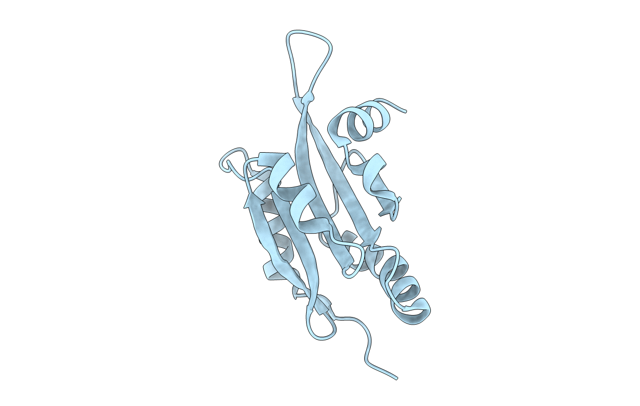
Title:
The Crystal Structure of Human Coactosin-like Protein at 1.9 A Resolution
Biological Source:
Source Organism(s):
Homo sapiens (Taxon ID: 9606)
Expression System(s):
Method Details:
Experimental Method:
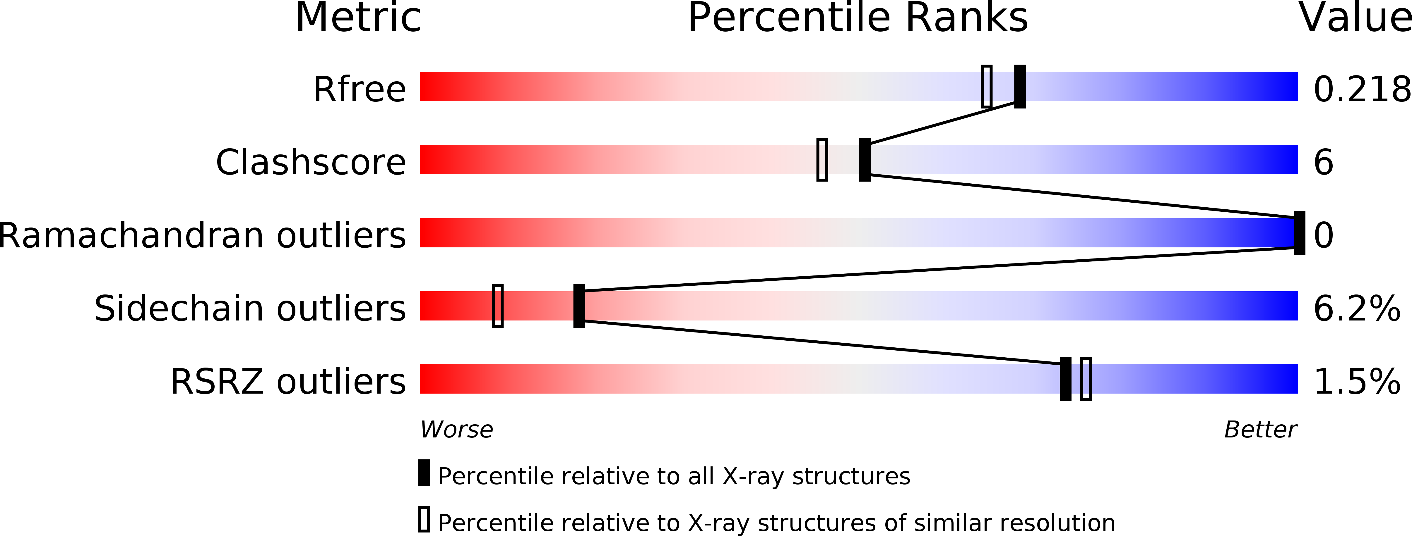
Resolution:
1.90 Å
R-Value Free:
0.21
R-Value Work:
0.16
R-Value Observed:
0.16
Space Group:
P 1 21 1