Deposition Date
2004-04-12
Release Date
2005-03-29
Last Version Date
2023-10-25
Entry Detail
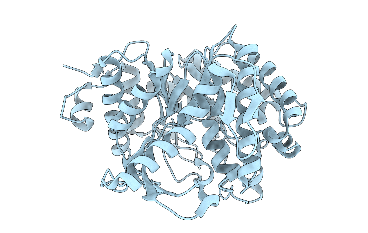
Biological Source:
Source Organism(s):
Pyrococcus horikoshii (Taxon ID: 53953)
Expression System(s):
Method Details:
Experimental Method:
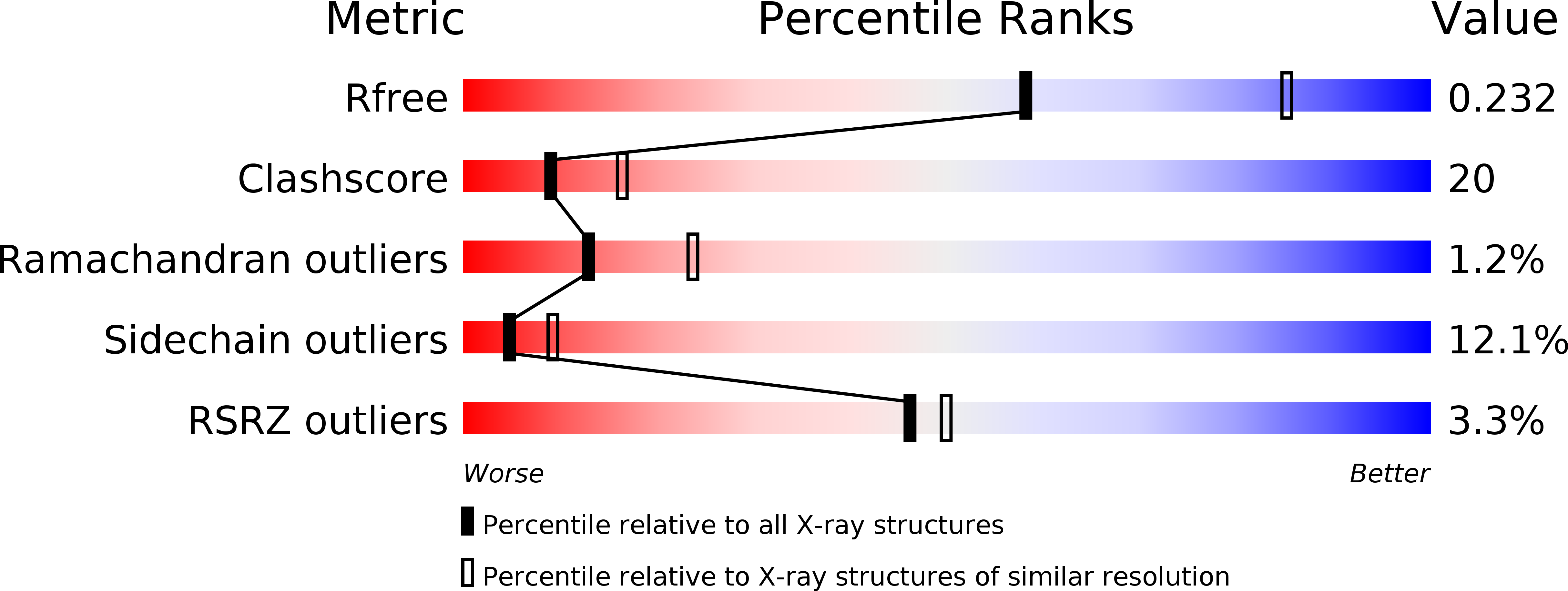
Resolution:
2.50 Å
R-Value Free:
0.23
R-Value Work:
0.17
R-Value Observed:
0.17
Space Group:
C 1 2 1