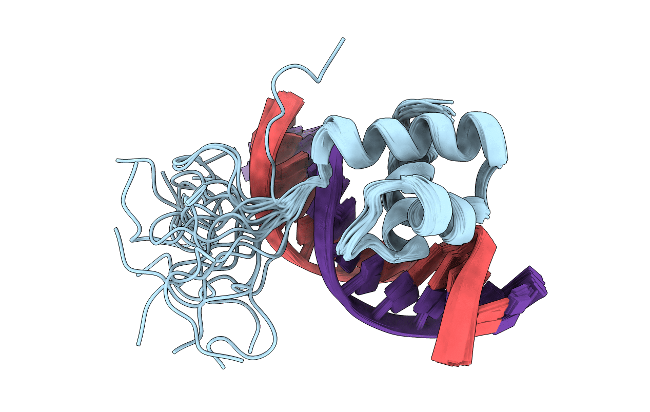
1VFC image
Deposition Date 2004-04-12
Release Date 2005-05-17
Last Version Date 2023-12-27
Entry Detail
PDB ID:
1VFC
Title:
Solution Structure Of The DNA Complex Of Human Trf2
Biological Source:
Source Organism:
Homo sapiens (Taxon ID: 9606)
(Taxon ID: )
Host Organism:
Method Details:
Experimental Method:
Conformers Calculated:
200
Conformers Submitted:
20
Selection Criteria:
structures with the lowest energy
Macromolecular Entities
Polymer Type:polypeptide(L)
Molecule:Telomeric repeat binding factor 2
Gene (Uniprot):TERF2
Chain IDs:C (auth: A)
Chain Length:63
Number of Molecules:1
Biological Source:Homo sapiens
Polymer Type:polydeoxyribonucleotide
Molecule:Short G-rich strand
Chain IDs:A (auth: B)
Chain Length:13
Number of Molecules:1
Biological Source:
Polymer Type:polydeoxyribonucleotide
Molecule:Short C-rich starnd
Chain IDs:B (auth: C)
Chain Length:13
Number of Molecules:1
Biological Source:
Ligand Molecules
Primary Citation
Comparison between TRF2 and TRF1 of their telomeric DNA-bound structures and DNA-binding activities
Protein Sci. 14 119 130 (2005)
PMID: 15608118 DOI: 10.1110/ps.04983705

Abstact

Mammalian telomeres consist of long tandem arrays of double-stranded telomeric TTAGGG repeats packaged by the telomeric DNA-binding proteins TRF1 and TRF2. Both contain a similar C-terminal Myb domain that mediates sequence-specific binding to telomeric DNA. In a DNA complex of TRF1, only the single Myb-like domain consisting of three helices can bind specifically to double-stranded telomeric DNA. TRF2 also binds to double-stranded telomeric DNA. Although the DNA binding mode of TRF2 is likely identical to that of TRF1, TRF2 plays an important role in the t-loop formation that protects the ends of telomeres. Here, to clarify the details of the double-stranded telomeric DNA-binding modes of TRF1 and TRF2, we determined the solution structure of the DNA-binding domain of human TRF2 bound to telomeric DNA; it consists of three helices, and like TRF1, the third helix recognizes TAGGG sequence in the major groove of DNA with the N-terminal arm locating in the minor groove. However, small but significant differences are observed; in contrast to the minor groove recognition of TRF1, in which an arginine residue recognizes the TT sequence, a lysine residue of TRF2 interacts with the TT part. We examined the telomeric DNA-binding activities of both DNA-binding domains of TRF1 and TRF2 and found that TRF1 binds more strongly than TRF2. Based on the structural differences of both domains, we created several mutants of the DNA-binding domain of TRF2 with stronger binding activities compared to the wild-type TRF2.

Legend

Protein

Chemical

Disease

Primary Citation of related structures