1VEM image
Deposition Date 2004-04-03
Release Date 2005-05-24
Last Version Date 2024-10-16
Entry Detail
PDB ID:
1VEM
Keywords:
Title:
Crystal Structure Analysis of Bacillus Cereus Beta-Amylase at the optimum pH (6.5)
Biological Source:
Source Organism:
Bacillus cereus (Taxon ID: 1396)
Host Organism:
Method Details:
Experimental Method:
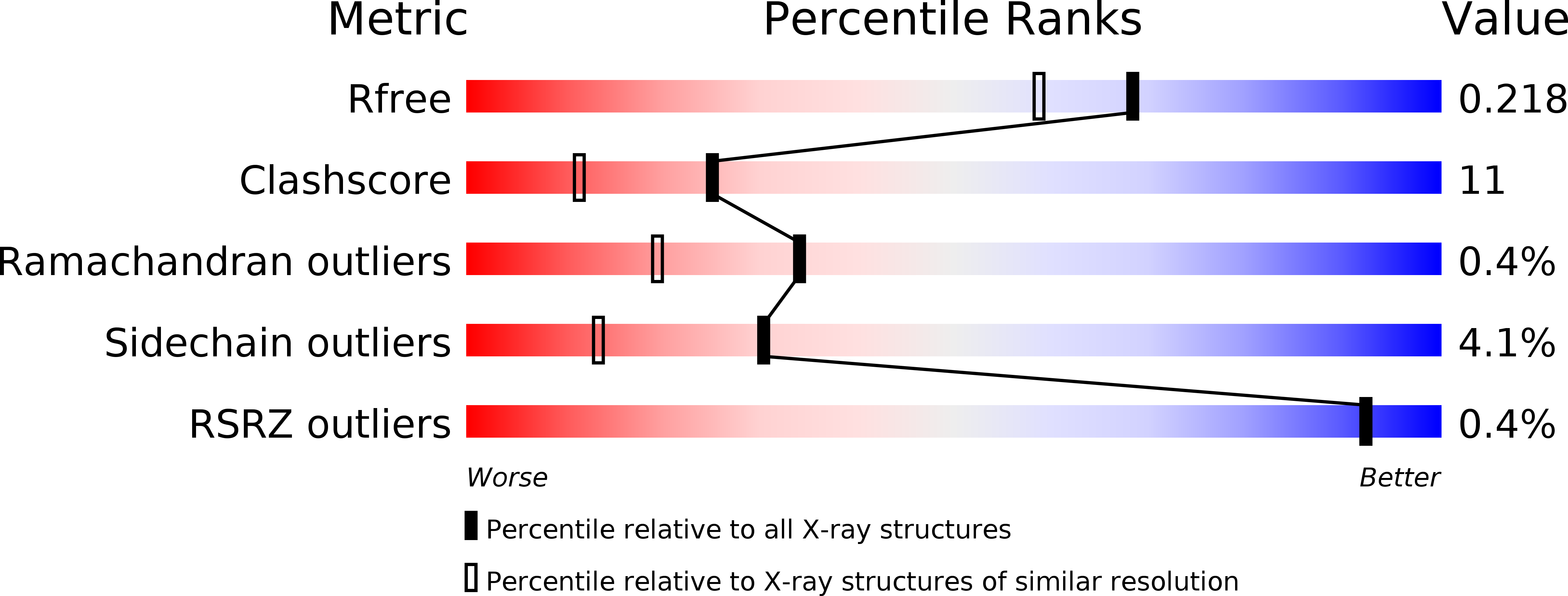
Resolution:
1.85 Å
R-Value Free:
0.20
R-Value Work:
0.17
R-Value Observed:
0.17
Space Group:
P 1 21 1
Macromolecular Entities
Polymer Type:polypeptide(L)
Molecule:Beta-amylase
Gene (Uniprot):spoII
Chain IDs:A
Chain Length:516
Number of Molecules:1
Biological Source:Bacillus cereus
Ligand Molecules
Peptide-like Molecules
PRD_900001
Primary Citation
Engineering of the pH optimum of Bacillus cereus beta-amylase: conversion of the pH optimum from a bacterial type to a higher-plant type
Biochemistry 43 12523 12531 (2004)
PMID: 15449941 DOI: 10.1021/bi049173h

Abstact

The optimum pH of Bacillus cereus beta-amylase (BCB, pH 6.7) differs from that of soybean beta-amylase (SBA, pH 5.4) due to the substitution of a few amino acid residues near the catalytic base residue (Glu 380 in SBA and Glu 367 in BCB). To explore the mechanism for controlling the optimum pH of beta-amylase, five mutants of BCB (Y164E, Y164F, Y164H, Y164Q, and Y164Q/T47M/Y164E/T328N) were constructed and characterized with respect to enzymatic properties and X-ray structural crystal analysis. The optimum pH of the four single mutants shifted to 4.2-4.8, approximately 2 pH units and approximately 1 pH unit lower than those of BCB and SBA, respectively, and their k(cat) values decreased to 41-3% of that of the wild-type enzyme. The X-ray crystal analysis of the enzyme-maltose complexes showed that Glu 367 of the wild type is surrounded by two water molecules (W1 and W2) that are not found in SBA. W1 is hydrogen-bonded to both side chains of Glu 367 and Tyr 164. The mutation of Tyr 164 to Glu and Phe resulted in the disruption of the hydrogen bond between Tyr 164 Oeta and W1 and the introduction of two additional water molecules near position 164. In contrast, the triple mutant of BCB with a slightly decreased pH optimum at pH 6.0 has no water molecules (W1 and W2) around Glu 367. These results suggested that a water-mediated hydrogen bond network (Glu 367...W1...Tyr 164...Thr 328) is the primary requisite for the increased pH optimum of wild-type BCB. This strategy is completely different from that of SBA, in which a hydrogen bond network (Glu 380...Thr 340...Glu 178) reduces the optimum pH in a hydrophobic environment.

Legend

Protein

Chemical

Disease

Primary Citation of related structures