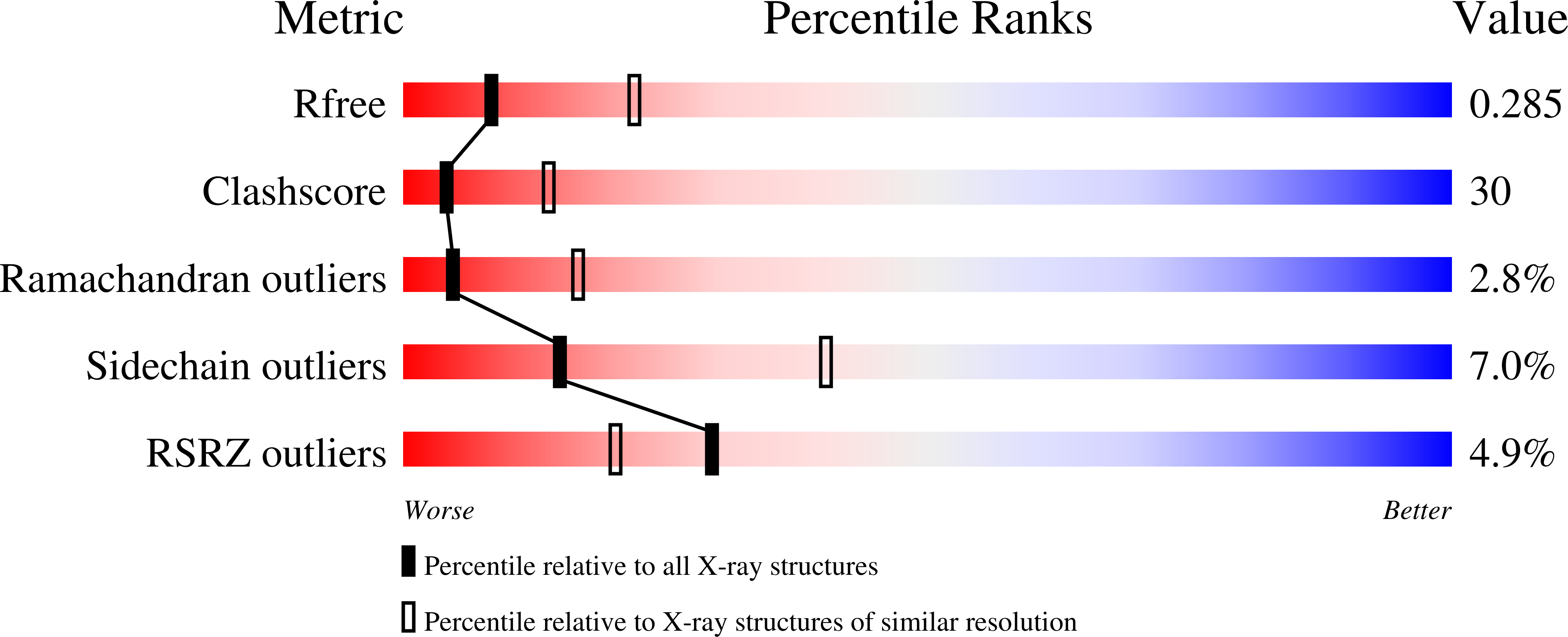
1VEA image
Deposition Date 2004-03-29
Release Date 2004-07-20
Last Version Date 2023-12-27
Entry Detail
PDB ID:
1VEA
Title:
Crystal Structure of HutP, an RNA binding antitermination protein
Biological Source:
Source Organism:
Method Details:
Experimental Method:
Resolution:
2.80 Å
R-Value Free:
0.28
R-Value Work:
0.23
R-Value Observed:
0.23
Space Group:
P 21 3
Macromolecular Entities
Polymer Type:polypeptide(L)
Molecule:Hut operon positive regulatory protein
Gene (Uniprot):hutP
Mutations:V51I
Chain IDs:A, B
Chain Length:148
Number of Molecules:2
Biological Source:Bacillus subtilis
Ligand Molecules
Primary Citation
Crystal Structure of Activated HutP; An RNA Binding Protein that Regulates Transcription of the hut Operon in Bacillus subtilis
Structure 12 1269 1280 (2004)
PMID: 15242603 DOI: 10.1016/j.str.2004.05.005

Abstact

HutP is an L-histidine-activated RNA binding protein that regulates the expression of the histidine utilization (hut) operon in Bacillus subtilis by binding to cis-acting regulatory sequences on the hut mRNA. The crystal structure of HutP complexed with an L-histidine analog showed a novel fold; there are four antiparallel beta strands in the central region of each monomer, with two alpha helices each on the front and back. Two HutP monomers form a dimer, and three dimers are arranged in crystallographic 3-fold symmetry to form a hexamer. A histidine analog was located in between the two monomers of HutP, with the imidazole group of L-histidine hydrogen bonded to Glu81. An activation mechanism is proposed based on the identification of key residues of HutP. The HutP binding region in hut mRNA was defined: it consists of three UAG trinucleotide motifs separated by four spacer nucleotides. Residues of HutP potentially important for RNA binding were identified.

Legend

Protein

Chemical

Disease

Primary Citation of related structures