Deposition Date
2004-03-27
Release Date
2004-11-02
Last Version Date
2024-10-16
Entry Detail
PDB ID:
1VE6
Keywords:
Title:
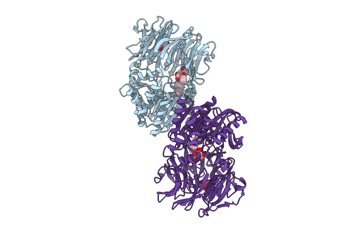
Crystal structure of an acylpeptide hydrolase/esterase from Aeropyrum pernix K1
Biological Source:
Source Organism(s):
Aeropyrum pernix (Taxon ID: 56636)
Expression System(s):
Method Details:
Experimental Method:
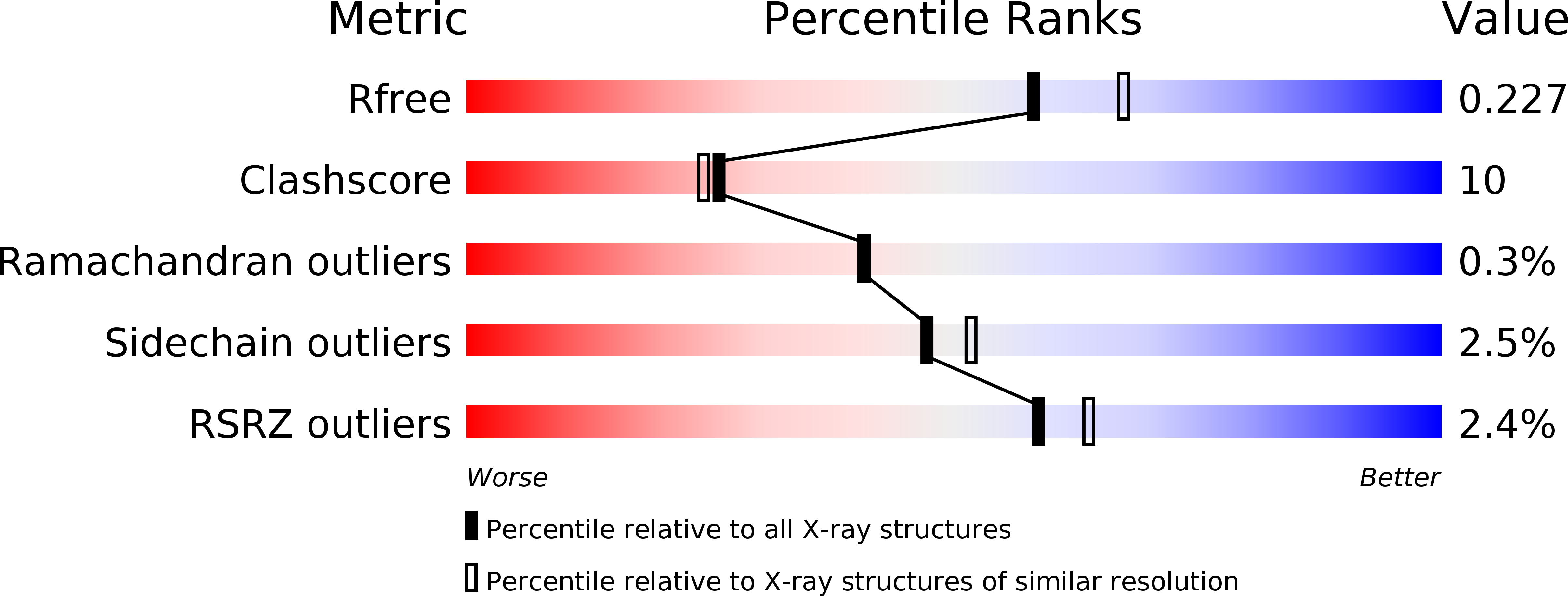
Resolution:
2.10 Å
R-Value Free:
0.22
R-Value Work:
0.19
R-Value Observed:
0.20
Space Group:
P 21 21 21