1VE3 image
Deposition Date 2004-03-26
Release Date 2005-05-24
Last Version Date 2024-10-30
Entry Detail
PDB ID:
1VE3
Title:
Crystal structure of PH0226 protein from Pyrococcus horikoshii OT3
Biological Source:
Source Organism:
Method Details:
Experimental Method:
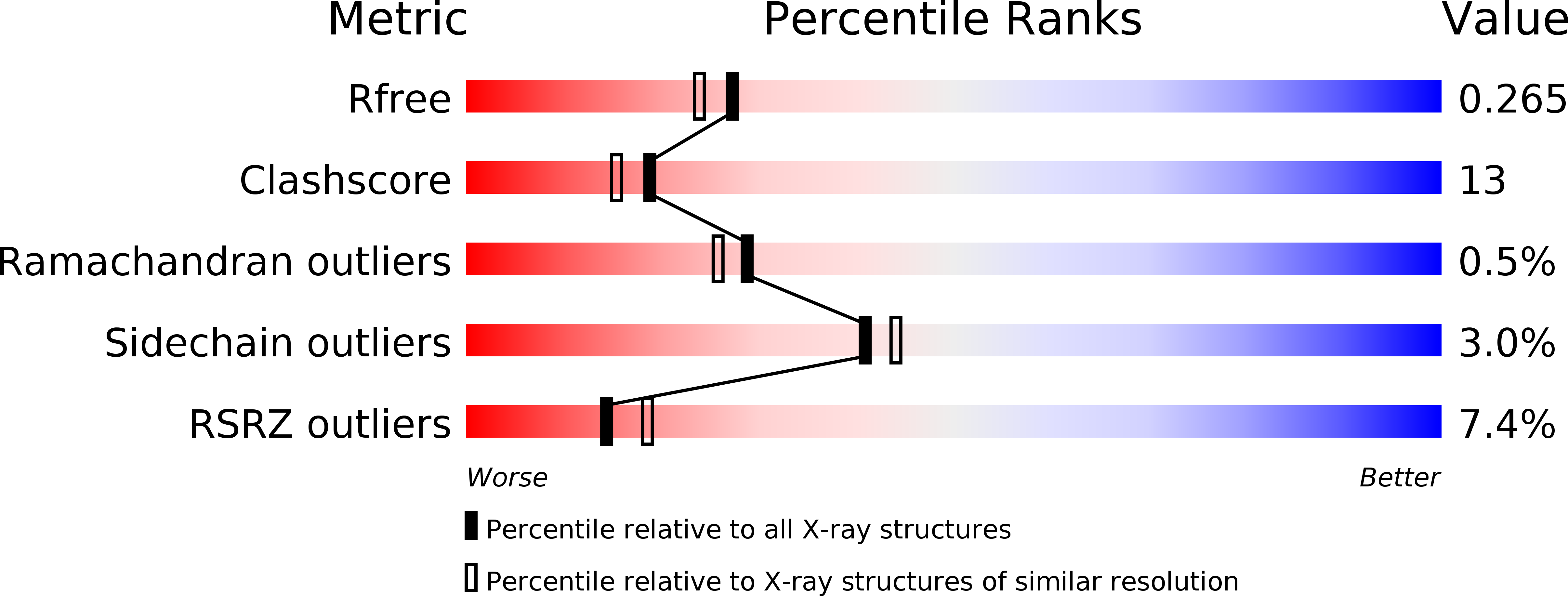
Resolution:
2.10 Å
R-Value Free:
0.27
R-Value Work:
0.23
R-Value Observed:
0.23
Space Group:
P 32 2 1
Macromolecular Entities
Structures with similar UniProt ID
Protein Blast
Polymer Type:polypeptide(L)
Molecule:hypothetical protein PH0226
Gene (Uniprot):PH0226
Chain IDs:A, B
Chain Length:227
Number of Molecules:2
Biological Source:Pyrococcus horikoshii
Modified Residue
Compound ID Chain ID Parent Comp ID Details 2D Image
MSE A MET SELENOMETHIONINE
Ligand Molecules
Primary Citation
Crystal structure of SAM-dependent methyltransferase from Pyrococcus horikoshii.
Acta Crystallogr.,Sect.F 73 706 712 (2017)
PMID: 29199993 DOI: 10.1107/S2053230X17016648

Abstact

Methyltransferases (MTs) are enzymes involved in methylation that are needed to perform cellular processes such as biosynthesis, metabolism, gene expression, protein trafficking and signal transduction. The cofactor S-adenosyl-L-methionine (SAM) is used for catalysis by SAM-dependent methyltransferases (SAM-MTs). The crystal structure of Pyrococcus horikoshii SAM-MT was determined to a resolution of 2.1 Å using X-ray diffraction. The monomeric structure consists of a Rossmann-like fold (domain I) and a substrate-binding domain (domain II). The cofactor (SAM) molecule binds at the interface between adjacent subunits, presumably near to the active site(s) of the enzyme. The observed dimeric state might be important for the catalytic function of the enzyme.

Legend

Protein

Chemical

Disease

Primary Citation of related structures