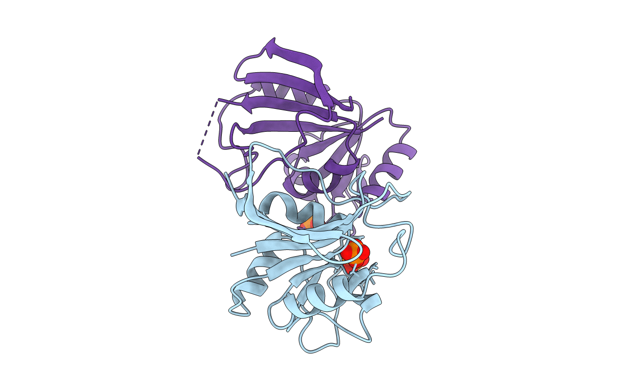
1VDR image
Deposition Date 1997-11-30
Release Date 1998-02-25
Last Version Date 2024-05-22
Entry Detail
PDB ID:
1VDR
Keywords:
Title:
DIHYDROFOLATE REDUCTASE
Biological Source:
Source Organism:
Method Details:
Experimental Method:
Resolution:
2.55 Å
R-Value Free:
0.30
R-Value Work:
0.18
R-Value Observed:
0.18
Space Group:
C 1 2 1
Macromolecular Entities
Polymer Type:polypeptide(L)
Molecule:DIHYDROFOLATE REDUCTASE
Gene (Uniprot):hdrA
Chain IDs:A, B
Chain Length:162
Number of Molecules:2
Biological Source:Haloferax volcanii
Ligand Molecules
Primary Citation
Structural features of halophilicity derived from the crystal structure of dihydrofolate reductase from the Dead Sea halophilic archaeon, Haloferax volcanii.
Structure 6 75 88 (1998)
PMID: 9493269 DOI: 10.1016/S0969-2126(98)00009-4

Abstact

BACKGROUND The proteins of halophilic archaea require high salt concentrations both for stability and for activity, whereas they denature at low ionic strength. The structural basis for this phenomenon is not yet well understood. The crystal structure of dihydrofolate reductase (DHFR) from Haloferax volcanii (hv-DHFR) reported here provides the third example of a structure of a protein from a halophilic organism. The enzyme is considered moderately halophilic, as it retains activity and secondary structure at monovalent salt concentrations as low as 0.5 M. RESULTS The crystal structure of hv-DHFR has been determined at 2.6 A resolution and reveals the same overall fold as that of other DHFRs. The structure is in the apo state, with an open conformation of the active-site gully different from the open conformation seen in other DHFR structures. The unique feature of hv-DHFR is a shift of the alpha helix encompassing residues 46-51 and an accompanied altered conformation of the ensuing loop relative to other DHFRs. Analysis of the charge distribution, amino acid composition, packing and hydrogen-bonding pattern in hv-DHFR and its non-halophilic homologs has been performed. CONCLUSIONS The moderately halophilic behavior of hv-DHFR is consistent with the lack of striking structural features expected to occur in extremely halophilic proteins. The most notable feature of halophilicity is the presence of clusters of non-interacting negatively charged residues. Such clusters are associated with unfavorable electrostatic energy at low salt concentrations, and may account for the instability of hv-DHFR at salt concentrations lower than 0.5 M. With respect to catalysis, the open conformation seen here is indicative of a conformational transition not reported previously. The impact of this conformation on function and/or halophilicity is unknown.

Legend

Protein

Chemical

Disease

Primary Citation of related structures