Deposition Date
2004-03-10
Release Date
2004-08-31
Last Version Date
2023-12-27
Entry Detail
PDB ID:
1VCO
Keywords:
Title:
Crystal Structure of T.th. HB8 CTP synthetase complex with Glutamine
Biological Source:
Source Organism:
Thermus thermophilus (Taxon ID: 274)
Host Organism:
Method Details:
Experimental Method:
Resolution:
2.15 Å
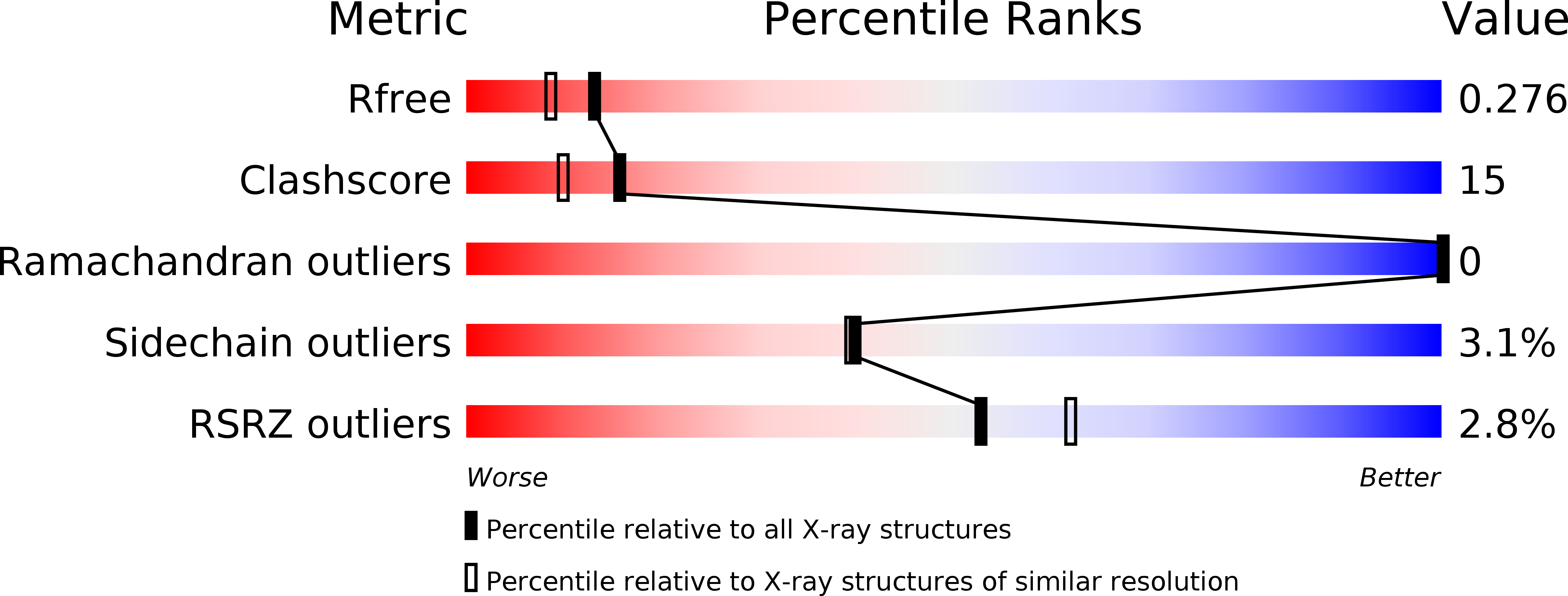
R-Value Free:
0.27
R-Value Work:
0.23
Space Group:
I 2 2 2