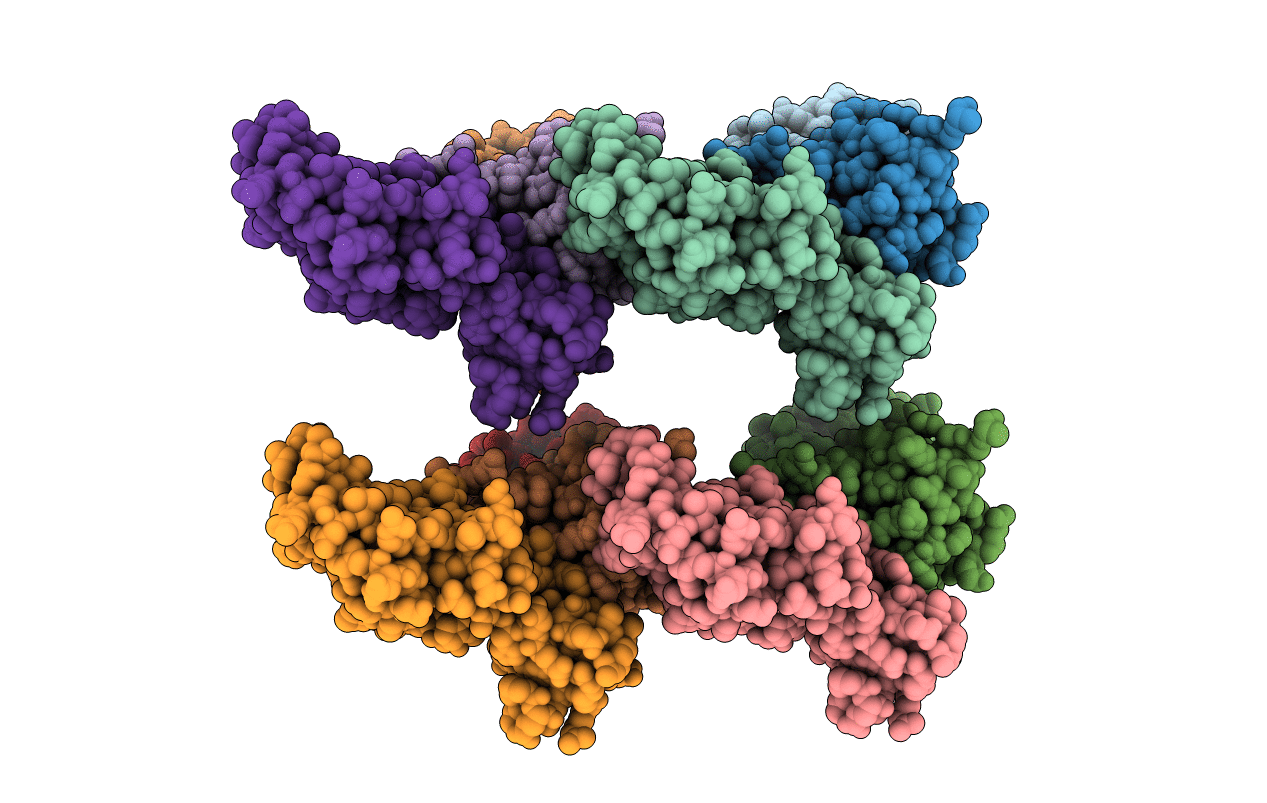
1VCB image
Deposition Date 1999-03-13
Release Date 1999-04-21
Last Version Date 2023-12-27
Entry Detail
PDB ID:
1VCB
Keywords:
Title:
THE VHL-ELONGINC-ELONGINB STRUCTURE
Biological Source:
Source Organism:
Homo sapiens (Taxon ID: 9606)
Method Details:
Experimental Method:
Resolution:
2.70 Å
R-Value Free:
0.28
R-Value Work:
0.23
Space Group:
P 41 2 2
Macromolecular Entities
Polymer Type:polypeptide(L)
Molecule:PROTEIN (ELONGIN B)
Gene (Uniprot):ELOB
Chain IDs:A, D, G, J
Chain Length:118
Number of Molecules:4
Biological Source:Homo sapiens
Polymer Type:polypeptide(L)
Molecule:PROTEIN (ELONGIN C)
Gene (Uniprot):ELOC
Chain IDs:B, E, H, K
Chain Length:112
Number of Molecules:4
Biological Source:Homo sapiens
Polymer Type:polypeptide(L)
Molecule:PROTEIN (VHL)
Gene (Uniprot):VHL
Chain IDs:C, F, I, L
Chain Length:160
Number of Molecules:4
Biological Source:Homo sapiens
Primary Citation
Structure of the VHL-ElonginC-ElonginB complex: implications for VHL tumor suppressor function.
Science 284 455 461 (1999)
PMID: 10205047 DOI: 10.1126/science.284.5413.455

Abstact

Mutation of the VHL tumor suppressor is associated with the inherited von Hippel-Lindau (VHL) cancer syndrome and the majority of kidney cancers. VHL binds the ElonginC-ElonginB complex and regulates levels of hypoxia-inducible proteins. The structure of the ternary complex at 2.7 angstrom resolution shows two interfaces, one between VHL and ElonginC and another between ElonginC and ElonginB. Tumorigenic mutations frequently occur in a 35-residue domain of VHL responsible for ElonginC binding. A mutational patch on a separate domain of VHL indicates a second macromolecular binding site. The structure extends the similarities to the SCF (Skp1-Cul1-F-box protein) complex that targets proteins for degradation, supporting the hypothesis that VHL may function in an analogous pathway.

Legend

Protein

Chemical

Disease

Primary Citation of related structures