1V9Y image
Deposition Date 2004-02-04
Release Date 2004-05-25
Last Version Date 2023-12-27
Entry Detail
PDB ID:
1V9Y
Title:
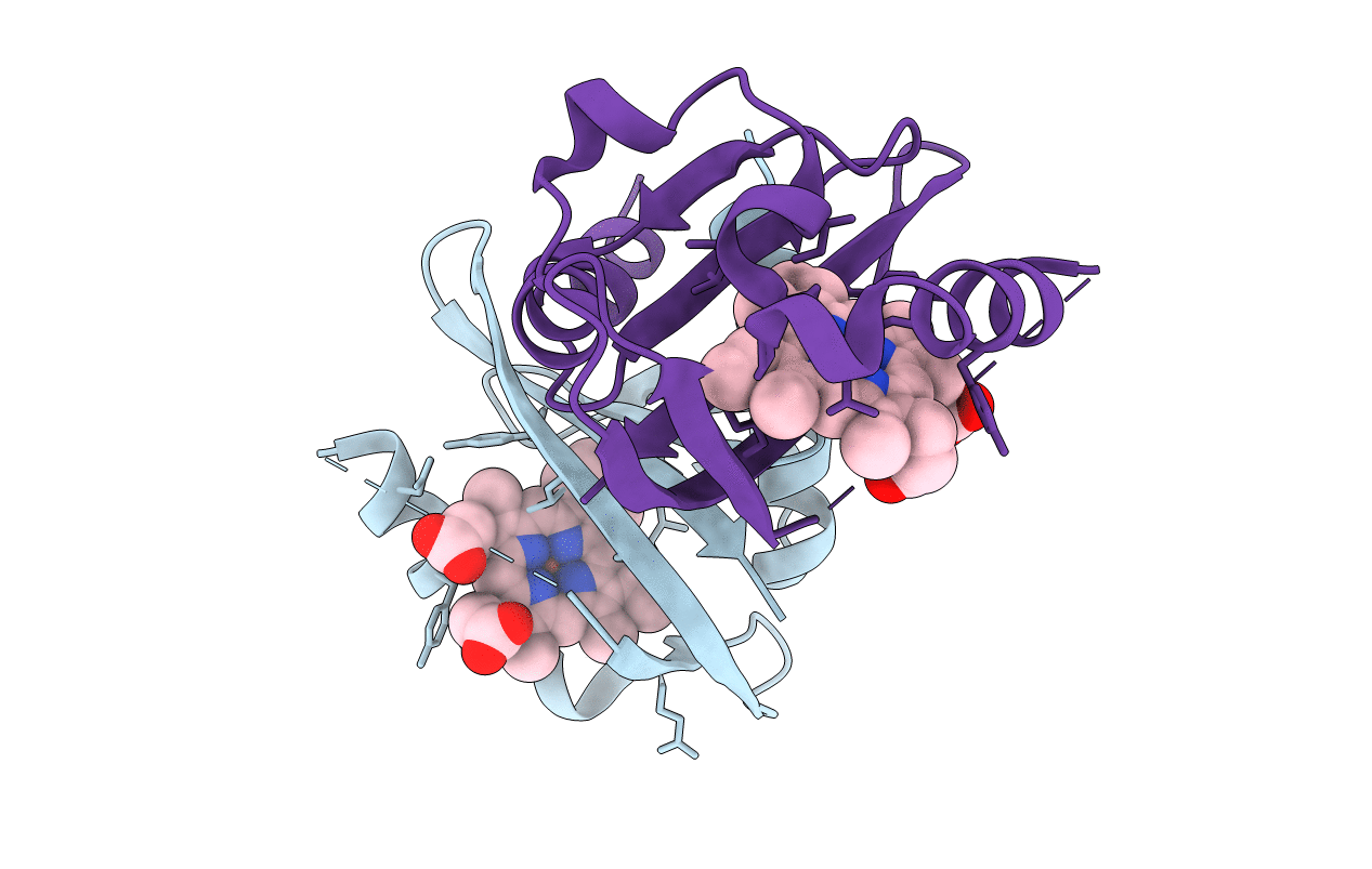
Crystal Structure of the heme PAS sensor domain of Ec DOS (ferric form)
Biological Source:
Source Organism:
Method Details:
Experimental Method:
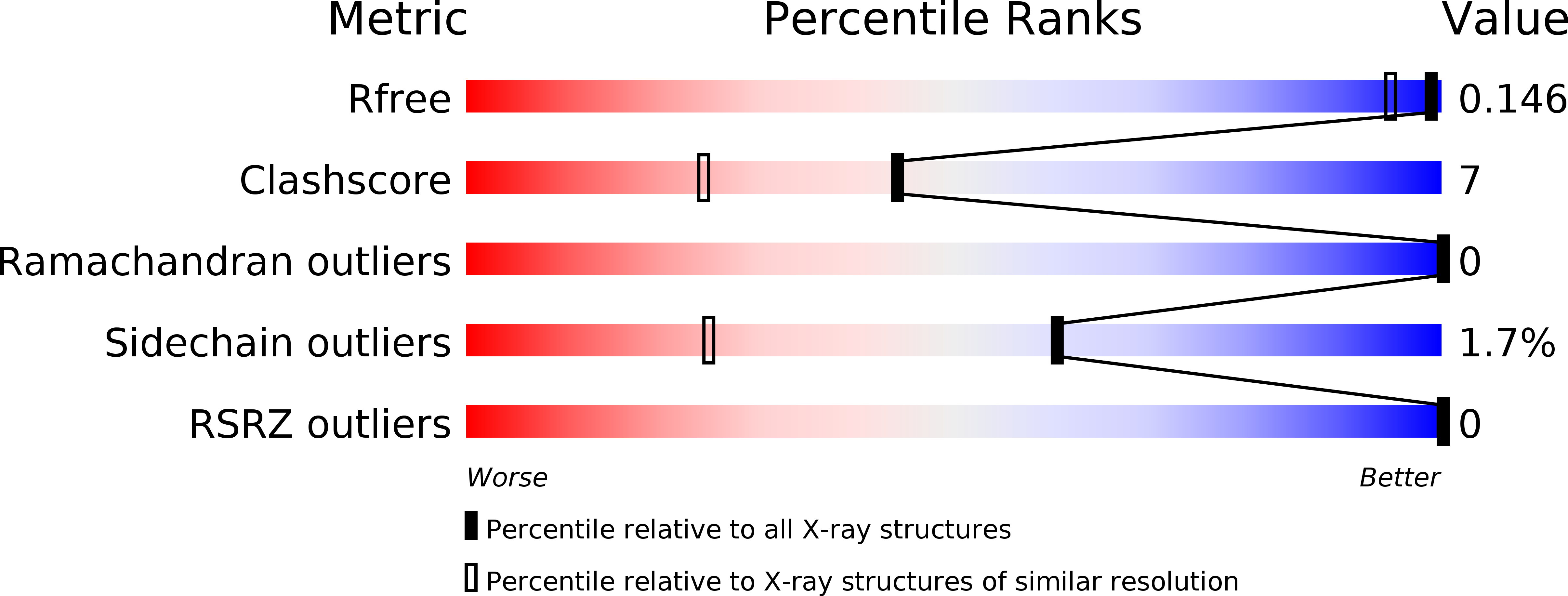
Resolution:
1.32 Å
R-Value Free:
0.21
R-Value Work:
0.16
R-Value Observed:
0.16
Space Group:
P 21 21 21
Macromolecular Entities
Polymer Type:polypeptide(L)
Molecule:Heme pas sensor protein
Chain IDs:A, B
Chain Length:167
Number of Molecules:2
Biological Source:Escherichia coli
Primary Citation
A redox-controlled molecular switch revealed by the crystal structure of a bacterial heme PAS sensor.
J.Biol.Chem. 279 20186 20193 (2004)
PMID: 14982921 DOI: 10.1074/jbc.M314199200

Abstact

PAS domains, which have been identified in over 1100 proteins from all three kingdoms of life, convert various input stimuli into signals that propagate to downstream components by modifying protein-protein interactions. One such protein is the Escherichia coli redox sensor, Ec DOS, a phosphodiesterase that degrades cyclic adenosine monophosphate in a redox-dependent manner. Here we report the crystal structures of the heme PAS domain of Ec DOS in both inactive Fe(3+) and active Fe(2+) forms at 1.32 and 1.9 A resolution, respectively. The protein folds into a characteristic PAS domain structure and forms a homodimer. In the Fe(3+) form, the heme iron is ligated to a His-77 side chain and a water molecule. Heme iron reduction is accompanied by heme-ligand switching from the water molecule to a side chain of Met-95 from the FG loop. Concomitantly, the flexible FG loop is significantly rigidified, along with a change in the hydrogen bonding pattern and rotation of subunits relative to each other. The present data led us to propose a novel redox-regulated molecular switch in which local heme-ligand switching may trigger a global "scissor-type" subunit movement that facilitates catalytic control.

Legend

Protein

Chemical

Disease

Primary Citation of related structures