
Deposition Date
2003-12-16
Release Date
2003-12-30
Last Version Date
2023-12-27
Entry Detail
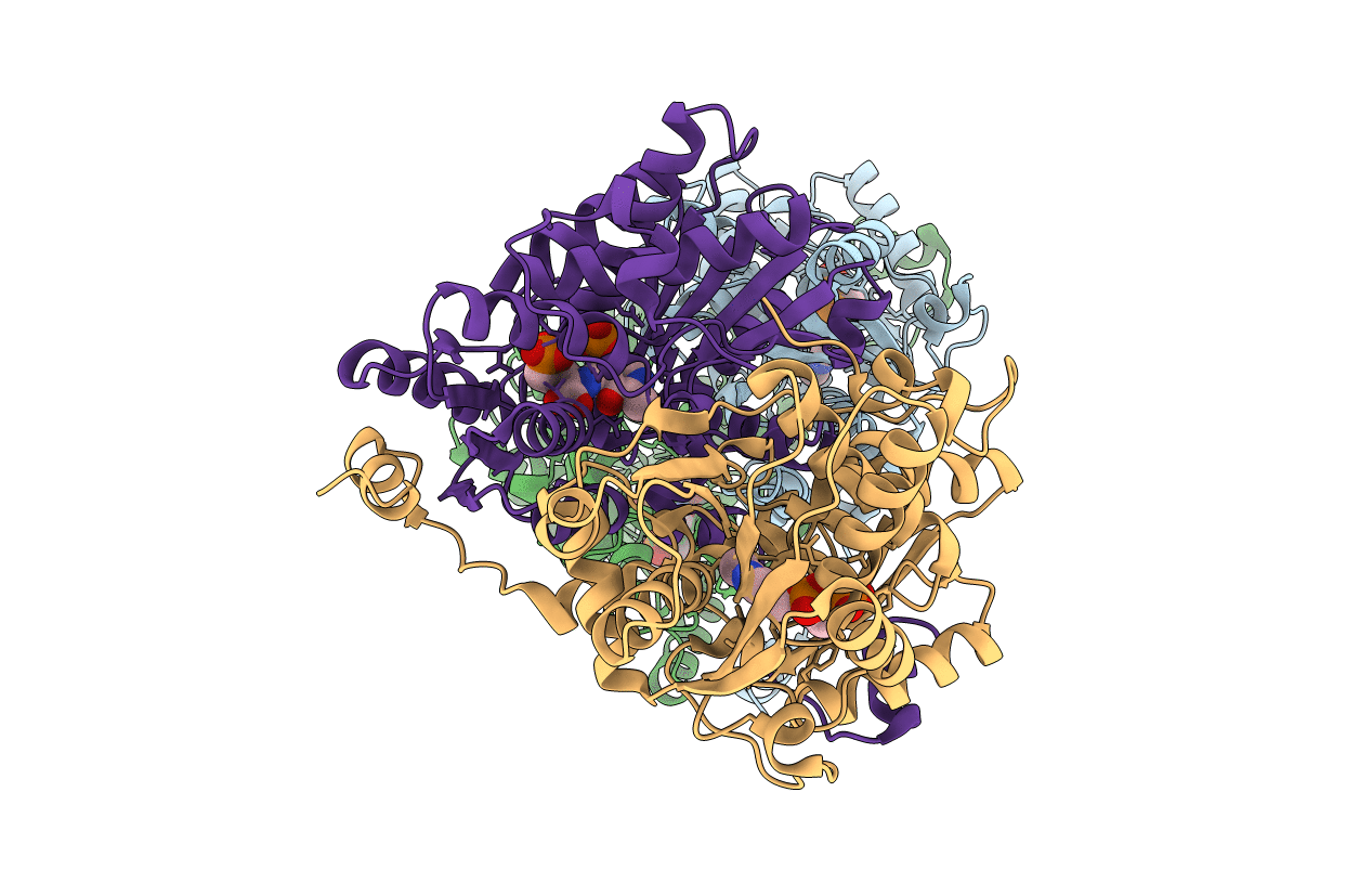
PDB ID:
1V7C
Keywords:
Title:
Crystal structure of threonine synthase from thermus thermophilus hb8 in complex with a substrate analogue
Biological Source:
Source Organism(s):
Thermus thermophilus (Taxon ID: 274)
Expression System(s):
Method Details:
Experimental Method:
Resolution:
2.00 Å
R-Value Free:
0.23
R-Value Work:
0.19
Space Group:
P 21 21 2
