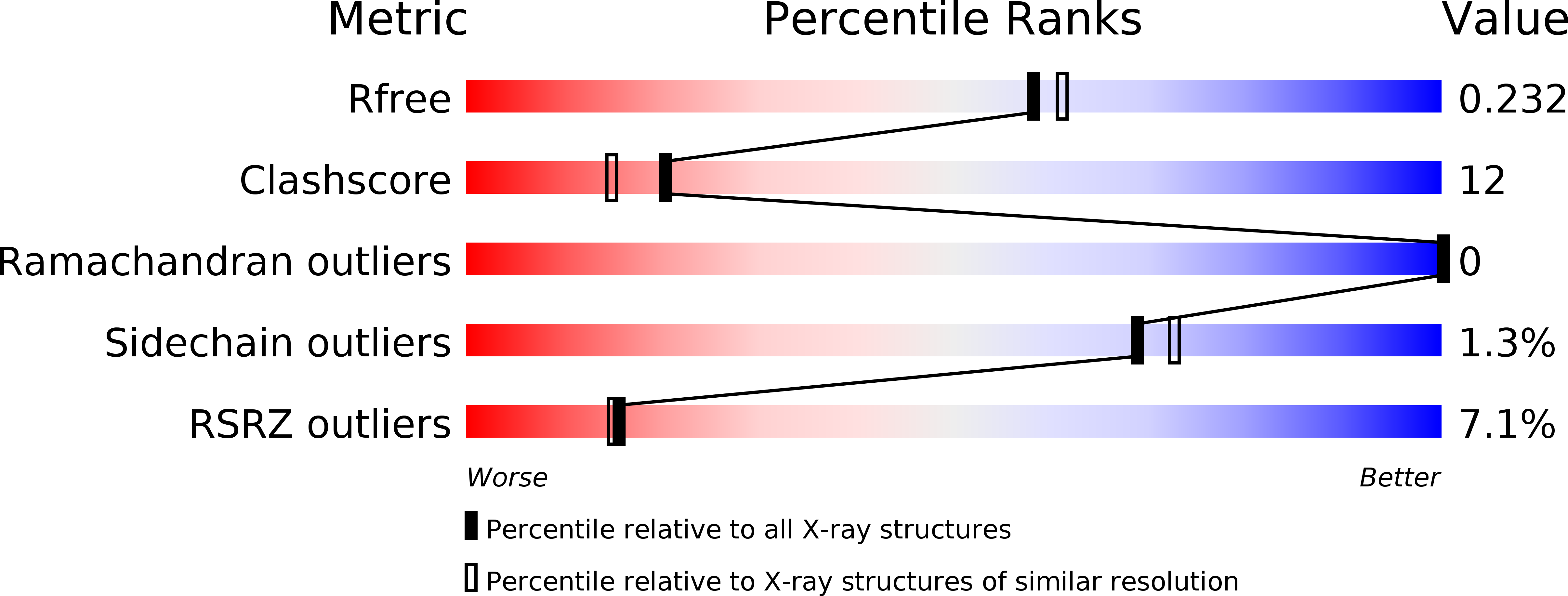
1V75 image
Deposition Date 2003-12-12
Release Date 2003-12-30
Last Version Date 2023-12-27
Entry Detail
PDB ID:
1V75
Title:
Crystal structure of hemoglobin D from the Aldabra giant tortoise (Geochelone gigantea) at 2.0 A resolution
Biological Source:
Source Organism:
Method Details:
Experimental Method:
Resolution:
2.02 Å
R-Value Free:
0.23
R-Value Work:
0.18
Space Group:
C 1 2 1
Macromolecular Entities
Structures with similar UniProt ID
Protein Blast
Polymer Type:polypeptide(L)
Molecule:Hemoglobin D alpha chain
Chain IDs:A
Chain Length:141
Number of Molecules:1
Biological Source:Dipsochelys dussumieri
Structures with similar UniProt ID
Protein Blast
Polymer Type:polypeptide(L)
Molecule:Hemoglobin A and D beta chain
Chain IDs:B
Chain Length:146
Number of Molecules:1
Biological Source:Dipsochelys dussumieri
Ligand Molecules
Primary Citation
Crystallization and preliminary X-ray diffraction study of hemoglobin D from the Aldabra giant tortoise, Geochelone gigantea.
Protein Pept.Lett. 10 422 425 (2003)
PMID: 14529497 DOI: 10.2174/0929866033478799

Abstact

Hemoglobin D (Hb D) from the Aldabra giant tortoise, Geochelone gigantea, was crystallized by the hanging drop vapor diffusion technique with a precipitant solution containing 10% polyethylene glycol 3350 and 50 mM HEPES-Na, pH 7.5. The Hb D crystals of G. gigantea, which diffract to at least a 2.0 A resolution, belong to the monoclinic space group C2 with unit cell dimensions of a = 112.1 A, b = 62.4 A, c = 54.0 A, and beta = 110.3 degrees. One alphabeta dimer molecule of Hb D existed in an asymmetric unit, with a calculated value of Vm of 2.77 A(3)Da(-1).

Legend

Protein

Chemical

Disease

Primary Citation of related structures