1V6J image
Deposition Date 2003-12-01
Release Date 2004-02-10
Last Version Date 2023-12-27
Entry Detail
PDB ID:
1V6J
Title:
peanut lectin-lactose complex crystallized in orthorhombic form at acidic pH
Biological Source:
Source Organism:
Method Details:
Experimental Method:
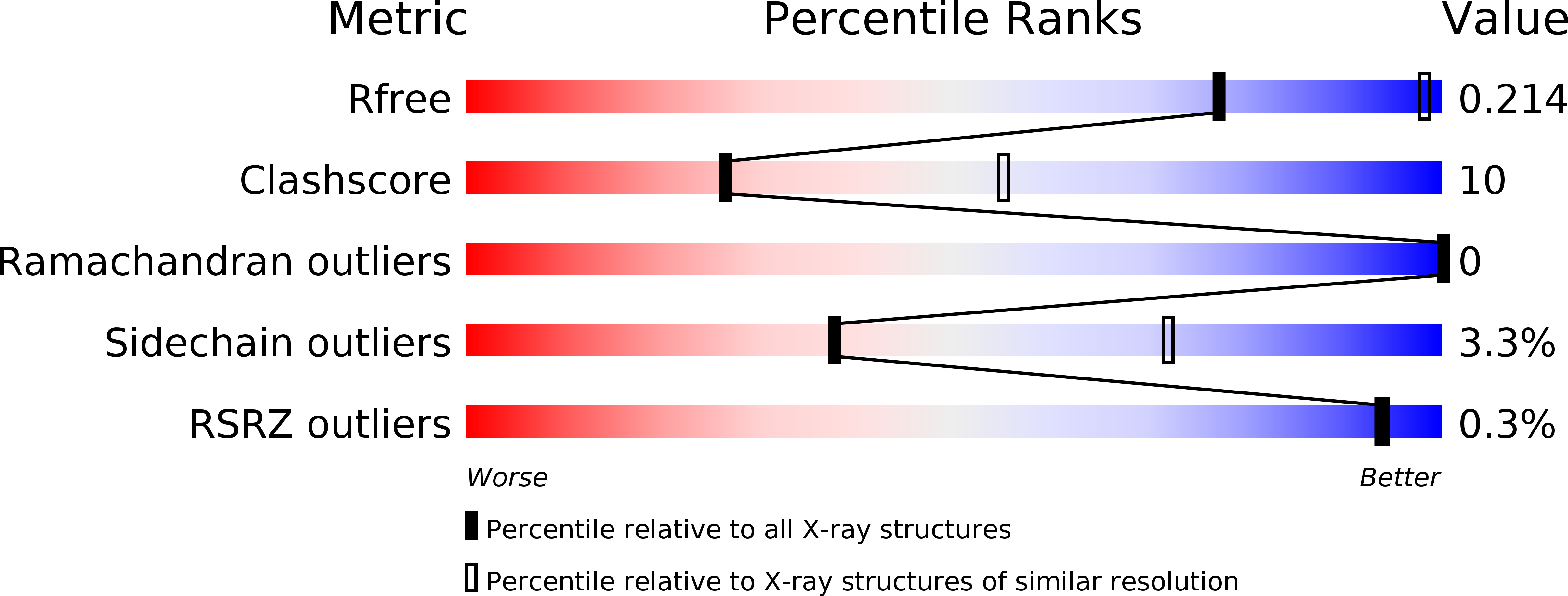
Resolution:
2.90 Å
R-Value Free:
0.22
R-Value Work:
0.16
Space Group:
P 21 21 2
Macromolecular Entities
Structures with similar UniProt ID
Protein Blast
Polymer Type:polypeptide(L)
Molecule:Galactose-binding lectin
Chain IDs:A, B, C, D
Chain Length:232
Number of Molecules:4
Biological Source:Arachis hypogaea
Peptide-like Molecules
PRD_900008
Primary Citation
Structural plasticity of peanut lectin: an X-ray analysis involving variation in pH, ligand binding and crystal structure.
Acta Crystallogr.,Sect.D 60 211 219 (2004)
PMID: 14747696 DOI: 10.1107/S090744490302849X

Abstact

Until recently, it has only been possible to grow crystals of peanut lectin when complexed with sugar ligands. It is now shown that it is possible to grow peanut lectin crystals at acidic pH in the presence of oligopeptides corresponding to a loop in the lectin molecule. Crystals have also been prepared in the presence of these peptides as well as lactose. Low-pH crystal forms of the lectin-lactose complex similar to those obtained at neutral pH have also been grown. Thus, crystals of peanut lectin grown under different environmental conditions, at two pH values with and without sugar bound to the lectin, are now available. They have been used to explore the plasticity and hydration of the molecule. A detailed comparison between different structures shows that the lectin molecule is sturdy and that the effect of changes in pH, ligand binding and environment on it is small. The region involving the curved front beta-sheet and the loops around the second hydrophobic core is comparatively rigid. The back beta-sheet involved in quaternary association, which exhibits considerable variability, is substantially flexible, as is the sugar-binding region. The numbers of invariant water molecules in the hydration shell are small and they are mainly involved in metal coordination or in stabilizing unusual structural features. Small consistent movements occur in the combining site upon sugar binding, although the site is essentially preformed.

Legend

Protein

Chemical

Disease

Primary Citation of related structures