1V5H image
Deposition Date 2003-11-23
Release Date 2004-06-08
Last Version Date 2023-12-27
Entry Detail
PDB ID:
1V5H
Title:
Crystal Structure of Human Cytoglobin (Ferric Form)
Biological Source:
Source Organism:
Homo sapiens (Taxon ID: 9606)
Method Details:
Experimental Method:
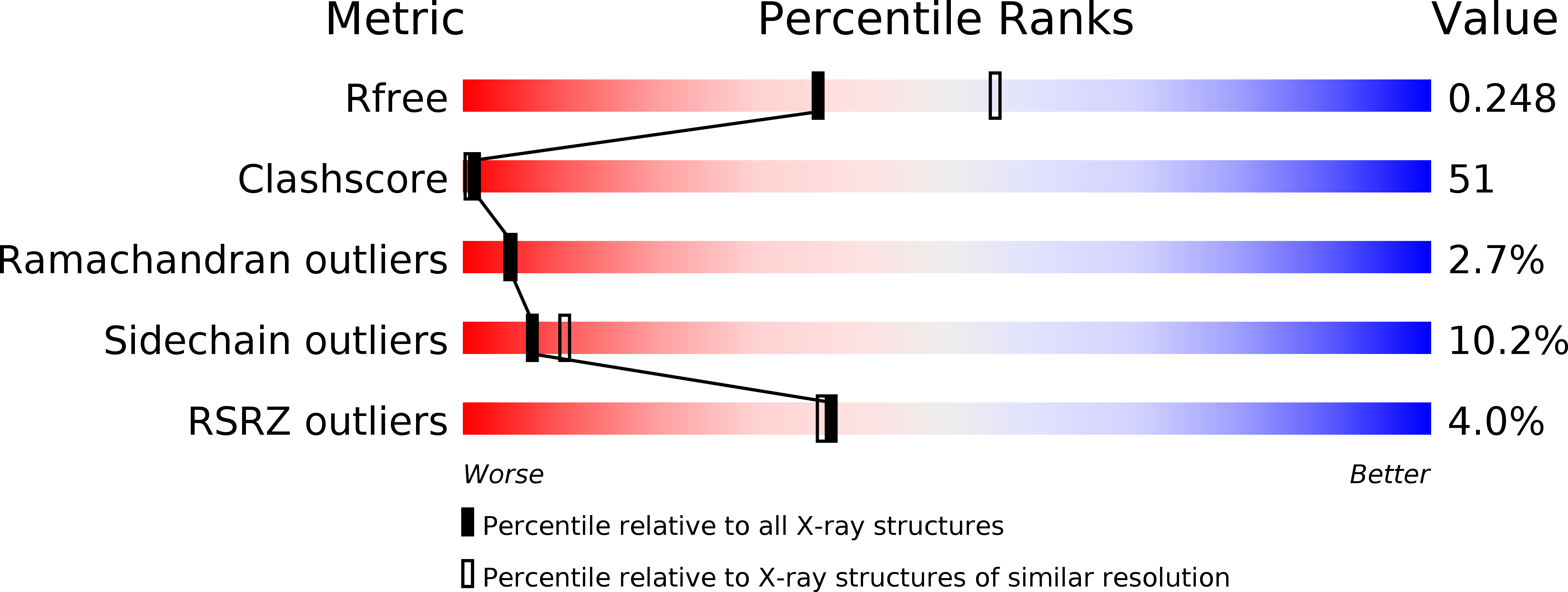
Resolution:
2.40 Å
R-Value Free:
0.25
R-Value Work:
0.24
R-Value Observed:
0.24
Space Group:
P 32 2 1
Macromolecular Entities
Structures with similar UniProt ID
Protein Blast
Polymer Type:polypeptide(L)
Molecule:Cytoglobin
Gene (Uniprot):CYGB
Chain IDs:A
Chain Length:193
Number of Molecules:1
Biological Source:Homo sapiens
Ligand Molecules
Primary Citation
Structural basis of human cytoglobin for ligand binding.
J.Mol.Biol. 339 873 885 (2004)
PMID: 15165856 DOI: 10.1016/j.jmb.2004.04.024

Abstact

Cytoglobin (Cgb), a newly discovered member of the vertebrate globin family, binds O(2) reversibly via its heme, as is the case for other mammalian globins (hemoglobin (Hb), myoglobin (Mb) and neuroglobin (Ngb)). While Cgb is expressed in various tissues, its physiological role is not clearly understood. Here, the X-ray crystal structure of wild type human Cgb in the ferric state at 2.4A resolution is reported. In the crystal structure, ferric Cgb is dimerized through two intermolecular disulfide bonds between Cys38(B2) and Cys83(E9), and the dimerization interface is similar to that of lamprey Hb and Ngb. The overall backbone structure of the Cgb monomer exhibits a traditional globin fold with a three-over-three alpha-helical sandwich, in which the arrangement of helices is basically the same among all globins studied to date. A detailed comparison reveals that the backbone structure of the CD corner to D helix region, the N terminus of the E-helix and the F-helix of Cgb resembles more closely those of pentacoordinated globins (Mb, lamprey Hb), rather than hexacoordinated globins (Ngb, rice Hb). However, the His81(E7) imidazole group coordinates directly to the heme iron as a sixth axial ligand to form a hexcoordinated heme, like Ngb and rice Hb. The position and orientation of the highly conserved residues in the heme pocket (Phe(CD1), Val(E11), distal His(E7) and proximal His(F8)) are similar to those of other globin proteins. Two alternative conformations of the Arg84(E10) guanidium group were observed, suggesting that it participates in ligand binding to Cgb, as is the case for Arg(E10) of Aplysia Mb and Lys(E10) of Ngb. The structural diversities and similarities among globin proteins are discussed with relevance to molecular evolutionary relationships.

Legend

Protein

Chemical

Disease

Primary Citation of related structures