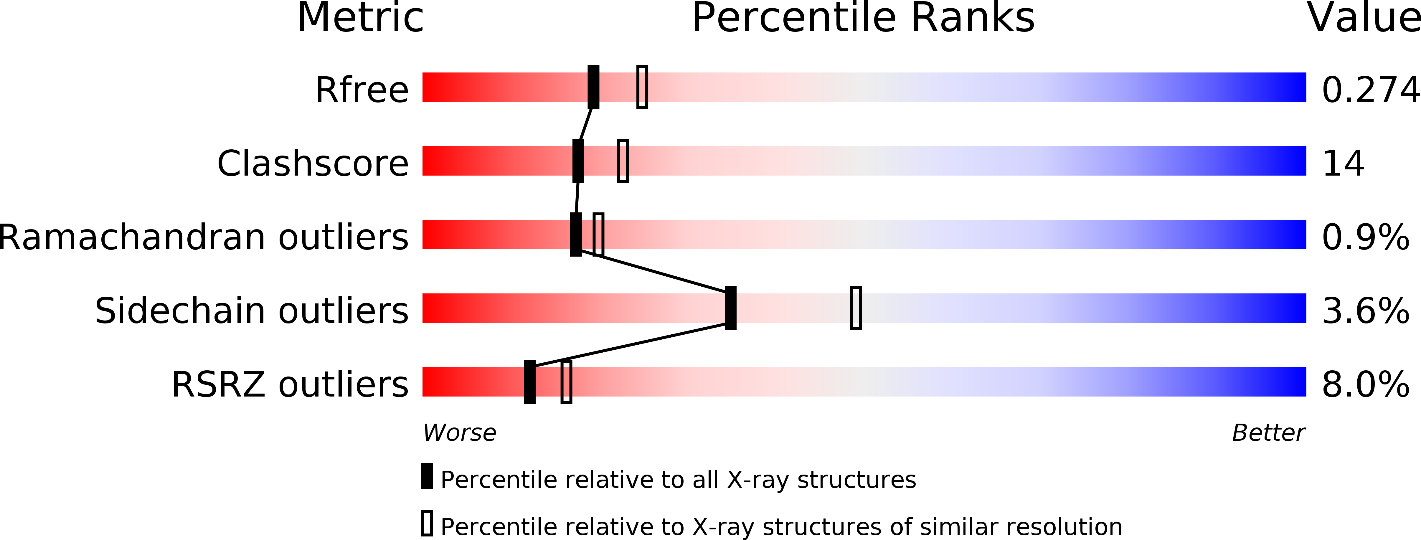
1V4S image
Deposition Date 2003-11-19
Release Date 2004-03-30
Last Version Date 2023-11-08
Entry Detail
PDB ID:
1V4S
Keywords:
Title:
Crystal structure of human glucokinase
Biological Source:
Source Organism:
Homo sapiens (Taxon ID: 9606)
Host Organism:
Method Details:
Experimental Method:
Resolution:
2.30 Å
R-Value Free:
0.27
R-Value Work:
0.23
R-Value Observed:
0.23
Space Group:
P 65 2 2
Macromolecular Entities
Polymer Type:polypeptide(L)
Molecule:glucokinase isoform 2
Gene (Uniprot):GCK
Chain IDs:A
Chain Length:455
Number of Molecules:1
Biological Source:Homo sapiens
Primary Citation
Structural basis for allosteric regulation of the monomeric allosteric enzyme human glucokinase
Structure 12 429 438 (2004)
PMID: 15016359 DOI: 10.1016/j.str.2004.02.005

Abstact

Glucokinase is a monomeric enzyme that displays a low affinity for glucose and a sigmoidal saturation curve for its substrate, two properties that are important for its playing the role of a glucose sensor in pancreas and liver. The molecular basis for these two properties is not well understood. Herein we report the crystal structures of glucokinase in its active and inactive forms, which demonstrate that global conformational change, including domain reorganization, is induced by glucose binding. This suggests that the positive cooperativity of monomeric glucokinase obeys the "mnemonical mechanism" rather than the well-known concerted model. These structures also revealed an allosteric site through which small molecules may modulate the kinetic properties of the enzyme. This finding provided the mechanistic basis for activation of glucokinase as a potential therapeutic approach for treating type 2 diabetes mellitus.

Legend

Protein

Chemical

Disease

Primary Citation of related structures