Deposition Date
2003-11-14
Release Date
2003-12-02
Last Version Date
2024-10-16
Entry Detail
PDB ID:
1V4L
Keywords:
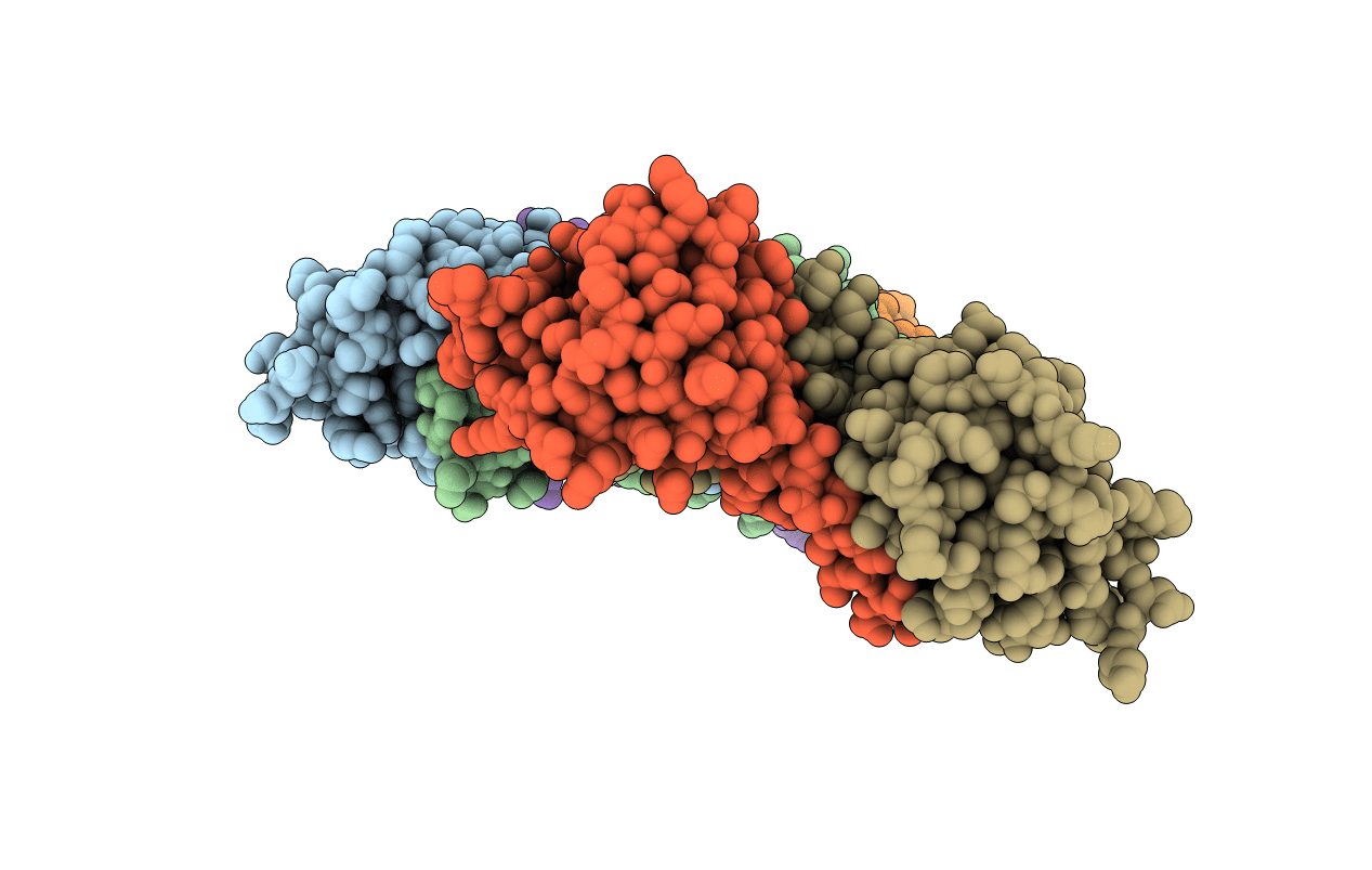
Title:
Crystal structure of a platelet agglutination factor isolated from the venom of Taiwan habu (Trimeresurus mucrosquamatus)
Biological Source:
Source Organism(s):
Protobothrops mucrosquamatus (Taxon ID: 103944)
Method Details:
Experimental Method:
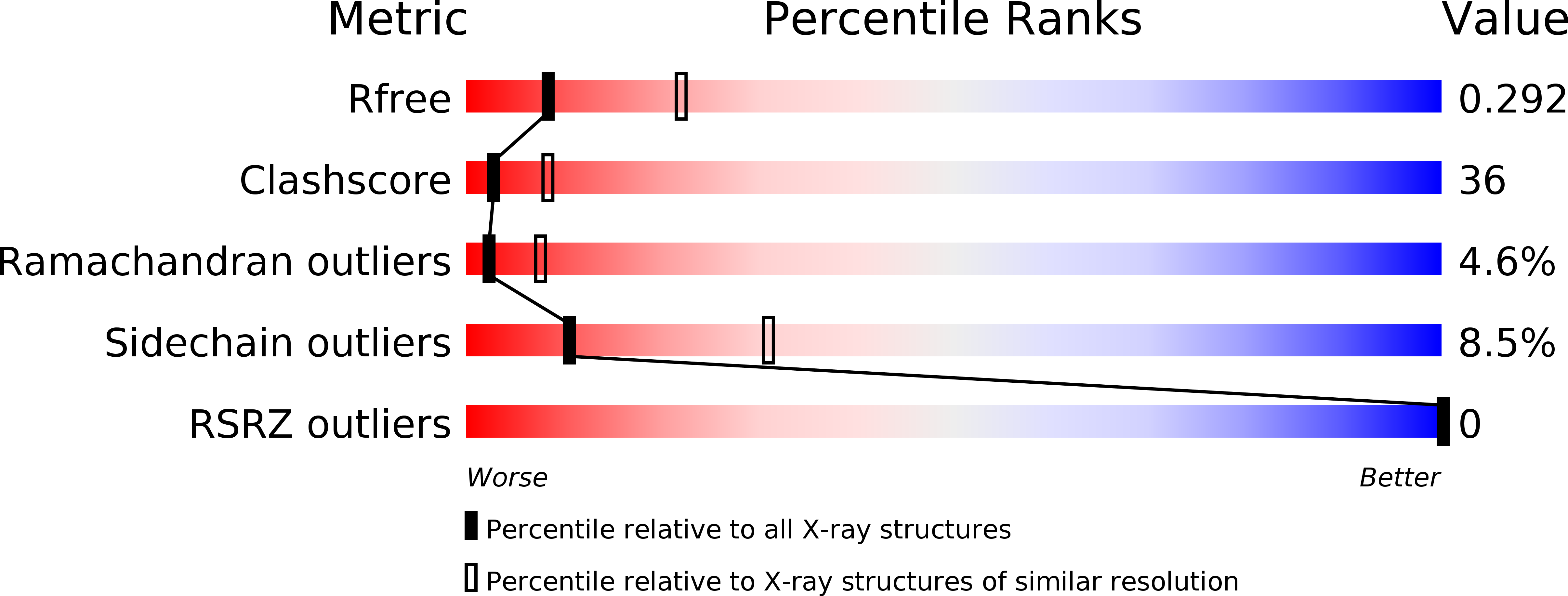
Resolution:
2.80 Å
R-Value Free:
0.29
R-Value Work:
0.23
Space Group:
I 4 2 2