Deposition Date
2003-11-13
Release Date
2004-10-05
Last Version Date
2024-10-30
Entry Detail
PDB ID:
1V4G
Keywords:
Title:
Crystal Structure of gamma-Glutamylcysteine Synthetase from Escherichia coli B
Biological Source:
Source Organism(s):
Escherichia coli (Taxon ID: 562)
Expression System(s):
Method Details:
Experimental Method:
Resolution:
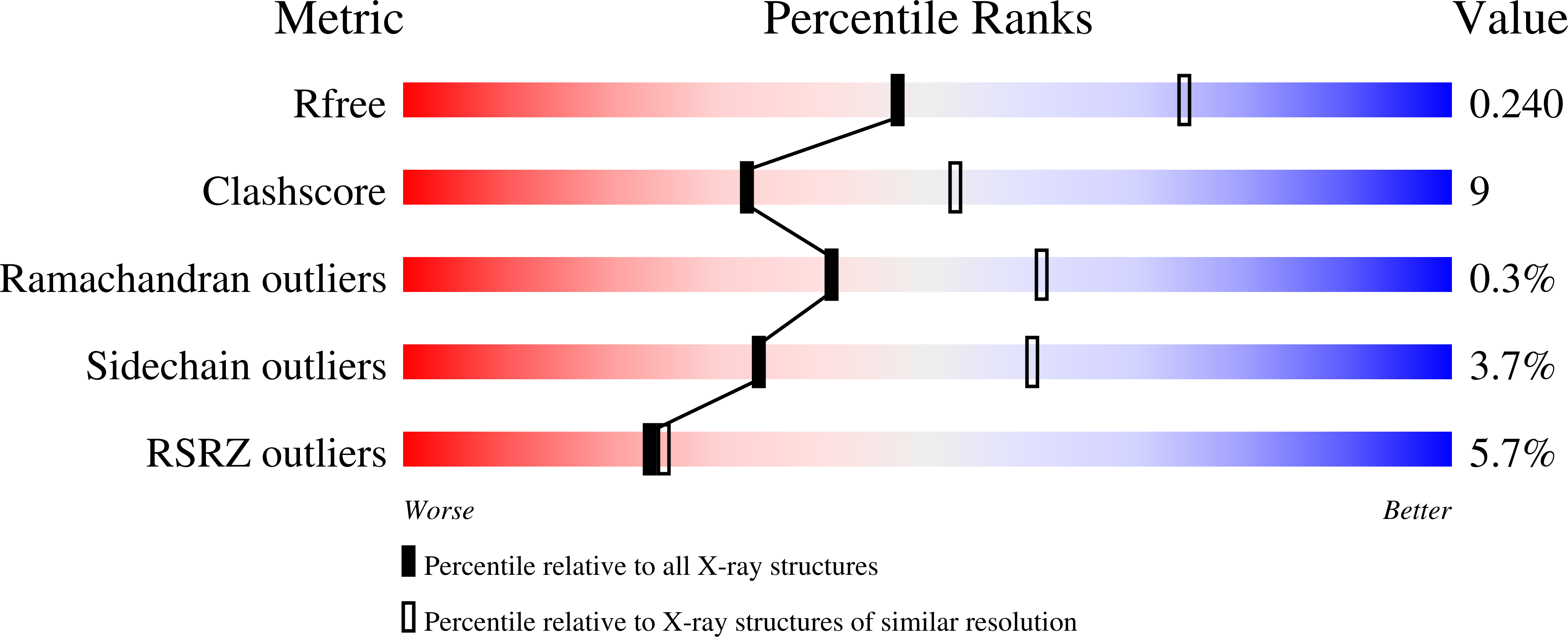
2.50 Å
R-Value Free:
0.23
R-Value Work:
0.20
R-Value Observed:
0.20
Space Group:
H 3