Deposition Date
2003-11-12
Release Date
2005-01-18
Last Version Date
2023-12-27
Entry Detail
PDB ID:
1V4B
Keywords:
Title:
The crystal structure of AzoR (Azo Reductase) from Escherichia coli: Oxidized form
Biological Source:
Source Organism(s):
Escherichia coli (Taxon ID: 562)
Expression System(s):
Method Details:
Experimental Method:
Resolution:
1.80 Å
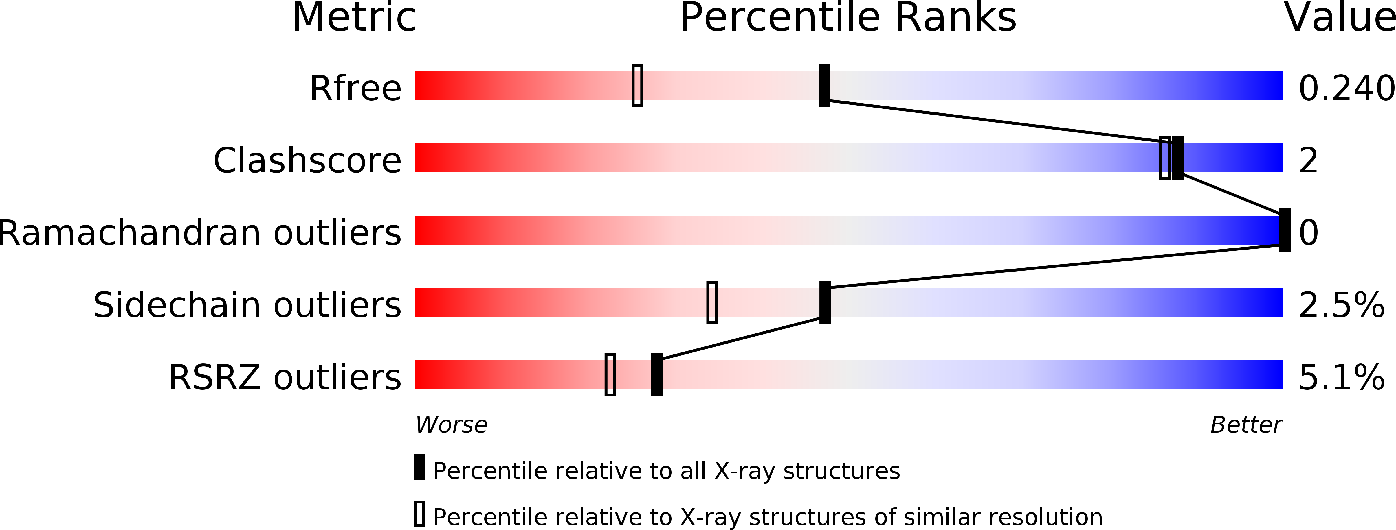
R-Value Free:
0.23
R-Value Work:
0.19
R-Value Observed:
0.19
Space Group:
P 42 21 2