1V3M image
Entry Detail
PDB ID:
1V3M
Keywords:
Title:
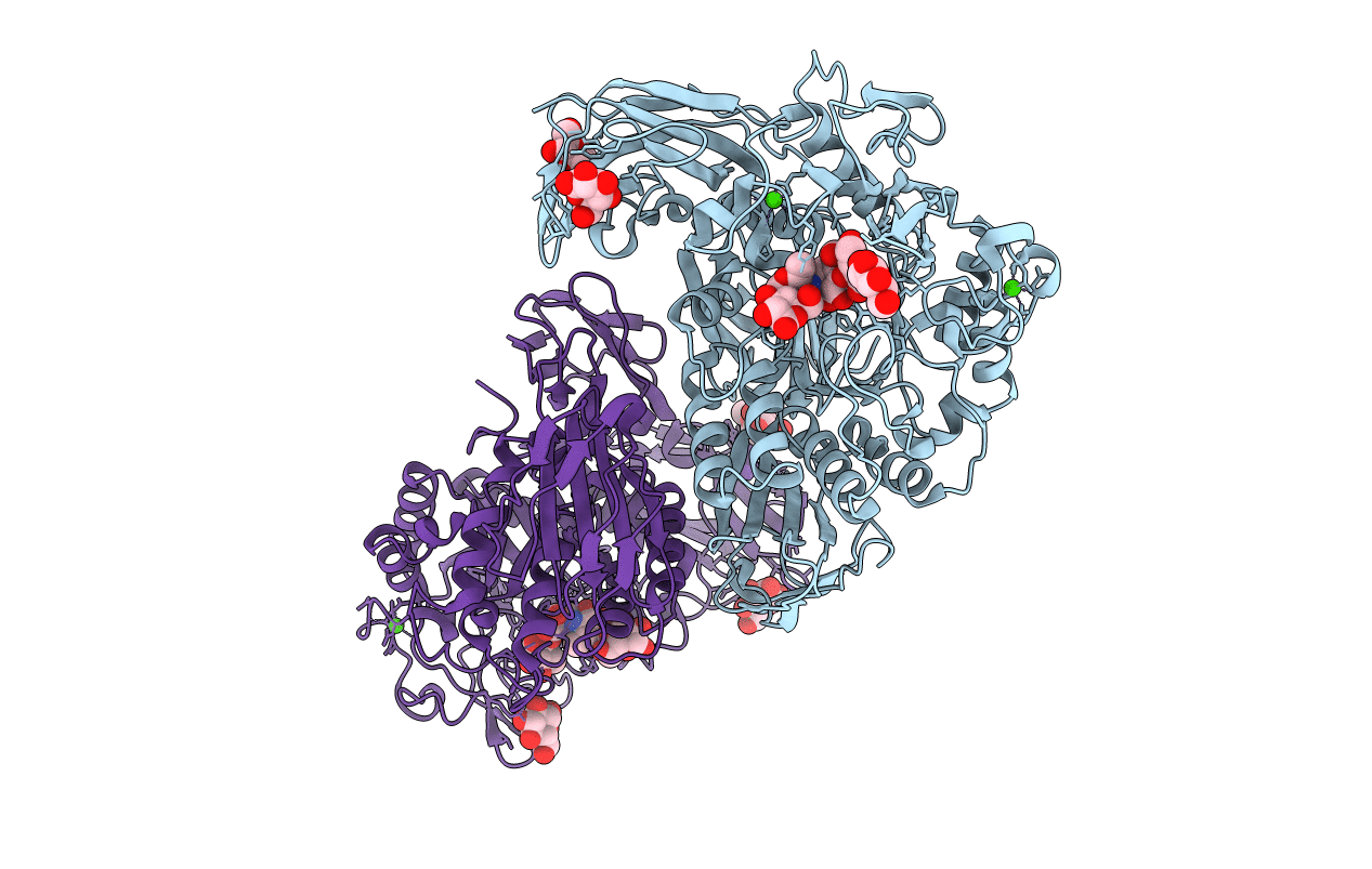
Crystal structure of F283Y mutant cyclodextrin glycosyltransferase complexed with a pseudo-tetraose derived from acarbose
Biological Source:
Source Organism:
Host Organism:
PDB Version:
Deposition Date:
2003-11-03
Release Date:
2004-08-03
Method Details:
Experimental Method:
Resolution:
2.00 Å
R-Value Free:
0.21
R-Value Work:
0.16
R-Value Observed:
0.16
Space Group:
P 1
Macromolecular Entities
Polymer Type:polypeptide(L)
Description:Cyclomaltodextrin glucanotransferase
Mutations:F283Y
Chain IDs:A, B
Chain Length:686
Number of Molecules:2
Biological Source:Bacillus sp.
Peptide-like Molecules
PRD_900001
Primary Citation
Role of Phe283 in enzymatic reaction of cyclodextrin glycosyltransferase from alkalophilic Bacillus sp.1011: Substrate binding and arrangement of the catalytic site
PROTEIN SCI. 13 457 465 (2004)
PMID: 14739329 DOI: 10.1110/ps.03408504

Abstact

Cyclodextrin glycosyltransferase (CGTase) belonging to the alpha-amylase family mainly catalyzes transglycosylation and produces cyclodextrins from starch and related alpha-1,4-glucans. The catalytic site of CGTase specifically conserves four aromatic residues, Phe183, Tyr195, Phe259, and Phe283, which are not found in alpha-amylase. To elucidate the structural role of Phe283, we determined the crystal structures of native and acarbose-complexed mutant CGTases in which Phe283 was replaced with leucine (F283L) or tyrosine (F283Y). The temperature factors of the region 259-269 in native F283L increased >10 A(2) compared with the wild type. The complex formation with acarbose not only increased the temperature factors (>10 A(2)) but also changed the structure of the region 257-267. This region is stabilized by interactions of Phe283 with Phe259 and Leu260 and plays an important role in the cyclodextrin binding. The conformation of the side-chains of Glu257, Phe259, His327, and Asp328 in the catalytic site was altered by the mutation of Phe283 with leucine, and this indicates that Phe283 partly arranges the structure of the catalytic site through contacts with Glu257 and Phe259. The replacement of Phe283 with tyrosine decreased the enzymatic activity in the basic pH range. The hydroxyl group of Tyr283 forms hydrogen bonds with the carboxyl group of Glu257, and the pK(a) of Glu257 in F283Y may be lower than that in the wild type.

Legend

Protein

Chemical

Disease

Primary Citation of related structures