
Deposition Date
2003-10-30
Release Date
2004-02-03
Last Version Date
2024-11-20
Entry Detail
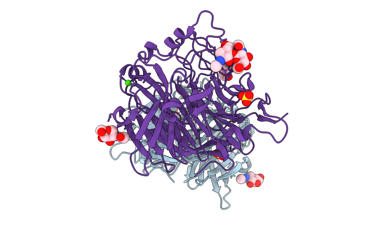
PDB ID:
1V3B
Keywords:
Title:
Structure of the hemagglutinin-neuraminidase from human parainfluenza virus type III
Biological Source:
Source Organism(s):
Human parainfluenza virus 3 (Taxon ID: 11216)
Expression System(s):
Method Details:
Experimental Method:
Resolution:
2.00 Å
R-Value Free:
0.21
R-Value Work:
0.18
R-Value Observed:
0.18
Space Group:
P 62 2 2
