1V2Z image
Deposition Date 2003-10-20
Release Date 2004-06-01
Last Version Date 2023-12-27
Entry Detail
PDB ID:
1V2Z
Title:
Crystal structure of the C-terminal domain of Thermosynechococcus elongatus BP-1 KaiA
Biological Source:
Source Organism:
Host Organism:
Method Details:
Experimental Method:
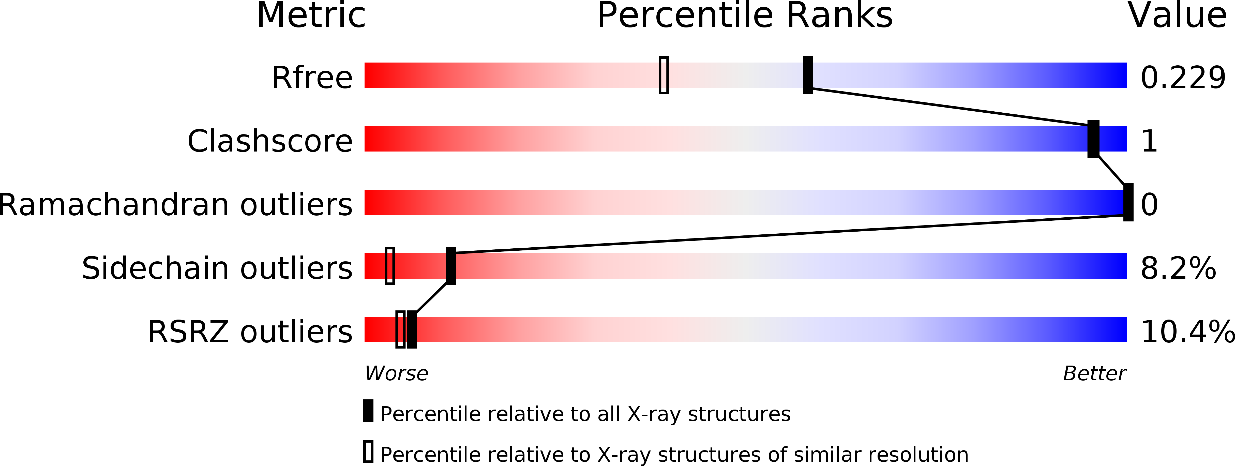
Resolution:
1.80 Å
R-Value Free:
0.26
R-Value Work:
0.22
R-Value Observed:
0.22
Space Group:
P 43 21 2
Macromolecular Entities
Polymer Type:polypeptide(L)
Molecule:circadian clock protein KaiA homolog
Chain IDs:A
Chain Length:111
Number of Molecules:1
Biological Source:Thermosynechococcus elongatus
Primary Citation
Crystal structure of the C-terminal clock-oscillator domain of the cyanobacterial KaiA protein
NAT.STRUCT.MOL.BIOL. 11 623 631 (2004)
PMID: 15170179 DOI: 10.1038/nsmb781

Abstact

KaiA, KaiB and KaiC constitute the circadian clock machinery in cyanobacteria, and KaiA activates kaiBC expression whereas KaiC represses it. Here we show that KaiA is composed of three functional domains, the N-terminal amplitude-amplifier domain, the central period-adjuster domain and the C-terminal clock-oscillator domain. The C-terminal domain is responsible for dimer formation, binding to KaiC, enhancing KaiC phosphorylation and generating the circadian oscillations. The X-ray crystal structure at a resolution of 1.8 A of the C-terminal clock-oscillator domain of KaiA from the thermophilic cyanobacterium Thermosynechococcus elongatus BP-1 shows that residue His270, located at the center of a KaiA dimer concavity, is essential to KaiA function. KaiA binding to KaiC probably occurs via the concave surface. On the basis of the structure, we predict the structural roles of the residues that affect circadian oscillations.

Legend

Protein

Chemical

Disease

Primary Citation of related structures