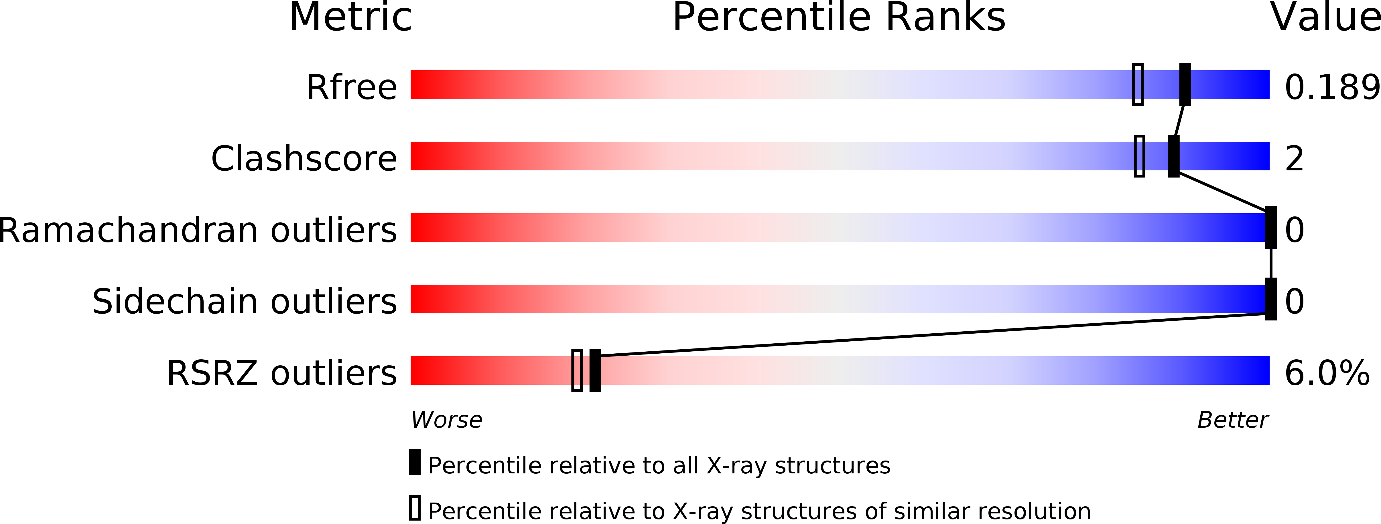
1V2B image
Entry Detail
PDB ID:
1V2B
Keywords:
Title:
Crystal Structure of PsbP Protein in the Oxygen-Evolving Complex of Photosystem II from Higher Plants
Biological Source:
Source Organism:
PDB Version:
Deposition Date:
2003-10-14
Release Date:
2004-05-18
Method Details:
Experimental Method:
Resolution:
1.60 Å
R-Value Free:
0.20
R-Value Work:
0.18
R-Value Observed:
0.18
Space Group:
P 21 21 2
Macromolecular Entities
Polymer Type:polypeptide(L)
Description:23-kDa polypeptide of photosystem II oxygen-evolving complex
Chain IDs:A, B
Chain Length:177
Number of Molecules:2
Biological Source:Nicotiana tabacum
Primary Citation
Crystal structure of the PsbP protein of photosystem II from Nicotiana tabacum
Embo Rep. 5 362 367 (2004)
PMID: 15031714 DOI: 10.1038/sj.embor.7400113

Abstact

PsbP is a membrane-extrinsic subunit of the water-oxidizing complex photosystem II (PS II). The evolutionary origin of PsbP has long been a mystery because it specifically exists in higher plants and green algae but not in cyanobacteria. We report here the crystal structure of PsbP from Nicotiana tabacum at a resolution of 1.6 A. Its structure is mainly composed of beta-sheet, and is not similar to any structures in cyanobacterial PS II. However, the electrostatic surface potential of PsbP is similar to that of cyanobacterial PsbV (cyt c(550)), which has a function similar to PsbP. A structural homology search with the DALI algorithm indicated that the folding of PsbP is very similar to that of Mog1p, a regulatory protein for the nuclear transport of Ran GTPase. The structure of PsbP provides insight into its novel function in GTP-regulated metabolism in PS II.

Legend

Protein

Chemical

Disease

Primary Citation of related structures