1V18 image
Deposition Date 2004-04-09
Release Date 2005-01-12
Last Version Date 2024-11-06
Entry Detail
PDB ID:
1V18
Title:
The crystal structure of beta-catenin armadillo repeat complexed with a phosphorylated APC 20mer repeat.
Biological Source:
Source Organism:
MUS MUSCULUS (Taxon ID: 10090)
HOMO SAPIENS (Taxon ID: 9606)
Host Organism:
Method Details:
Experimental Method:
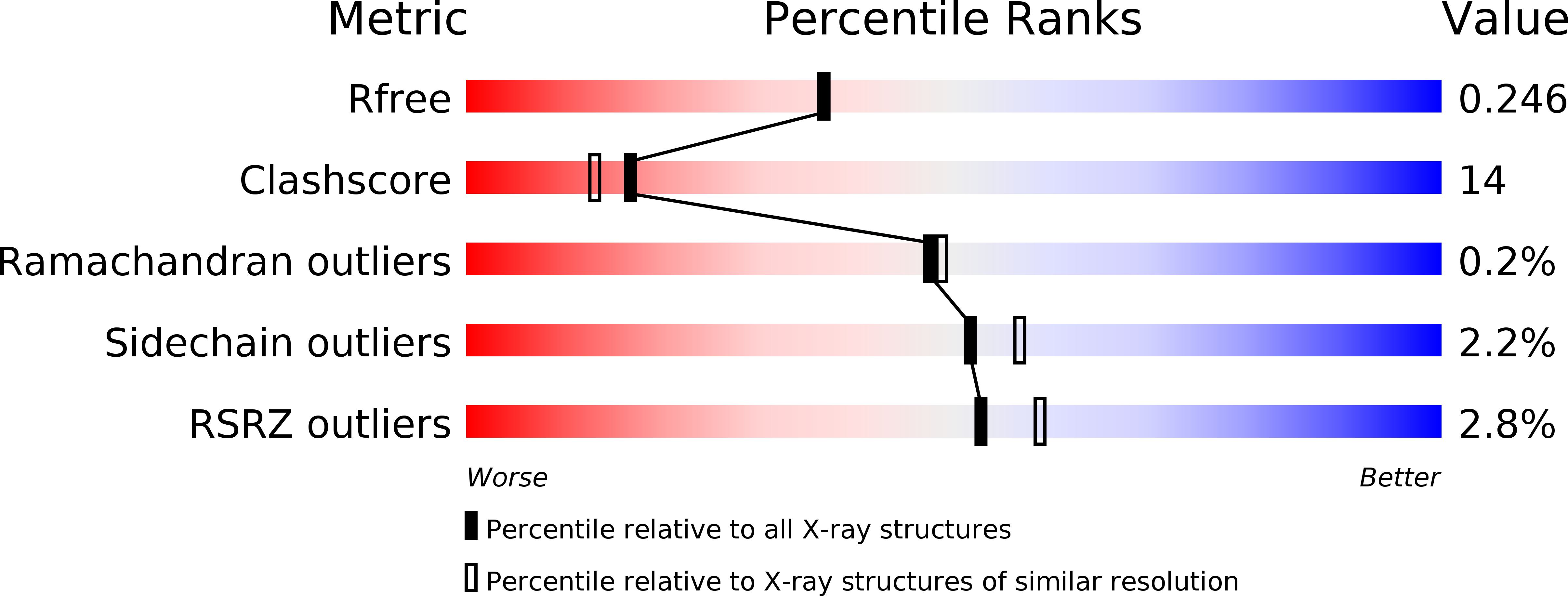
Resolution:
2.10 Å
R-Value Free:
0.25
R-Value Work:
0.20
R-Value Observed:
0.20
Space Group:
P 21 21 21
Macromolecular Entities
Structures with similar UniProt ID
Protein Blast
Polymer Type:polypeptide(L)
Molecule:BETA-CATENIN
Gene (Uniprot):Ctnnb1
Chain IDs:A
Chain Length:538
Number of Molecules:1
Biological Source:MUS MUSCULUS
Structures with similar UniProt ID
Protein Blast
Polymer Type:polypeptide(L)
Molecule:ADENOMATOUS POLYPOSIS COLI
Gene (Uniprot):APC
Chain IDs:B
Chain Length:47
Number of Molecules:1
Biological Source:HOMO SAPIENS
Modified Residue
Compound ID Chain ID Parent Comp ID Details 2D Image
SEP B SER PHOSPHOSERINE
Primary Citation
Mechanism of Phosphorylation-Dependent Binding of Apc to Beta-Catenin and its Role in Beta-Catenin Degradation
Mol.Cell 15 511 ? (2004)
PMID: 15327768 DOI: 10.1016/J.MOLCEL.2004.08.010

Abstact

The transcriptional coactivator beta-catenin mediates Wnt growth factor signaling. In the absence of a Wnt signal, casein kinase 1 (CK1) and glycogen synthase kinase-3beta (GSK-3beta) phosphorylate cytosolic beta-catenin, thereby flagging it for recognition and destruction by the ubiquitin/proteosome machinery. Phosphorylation occurs in a multiprotein complex that includes the kinases, beta-catenin, axin, and the Adenomatous Polyposis Coli (APC) protein. The role of APC in this process is poorly understood. CK1epsilon and GSK-3beta phosphorylate APC, which increases its affinity for beta-catenin. Crystal structures of phosphorylated and nonphosphorylated APC bound to beta-catenin reveal a phosphorylation-dependent binding motif generated by mutual priming of CK1 and GSK-3beta substrate sequences. Axin is shown to act as a scaffold for substrate phosphorylation by these kinases. Phosphorylated APC and axin bind to the same surface of, and compete directly for, beta-catenin. The structural and biochemical data suggest a novel model for how APC functions in beta-catenin degradation.

Legend

Protein

Chemical

Disease

Primary Citation of related structures