1V0F image
Deposition Date 2004-03-28
Release Date 2004-12-13
Last Version Date 2024-05-08
Entry Detail
PDB ID:
1V0F
Keywords:
Title:
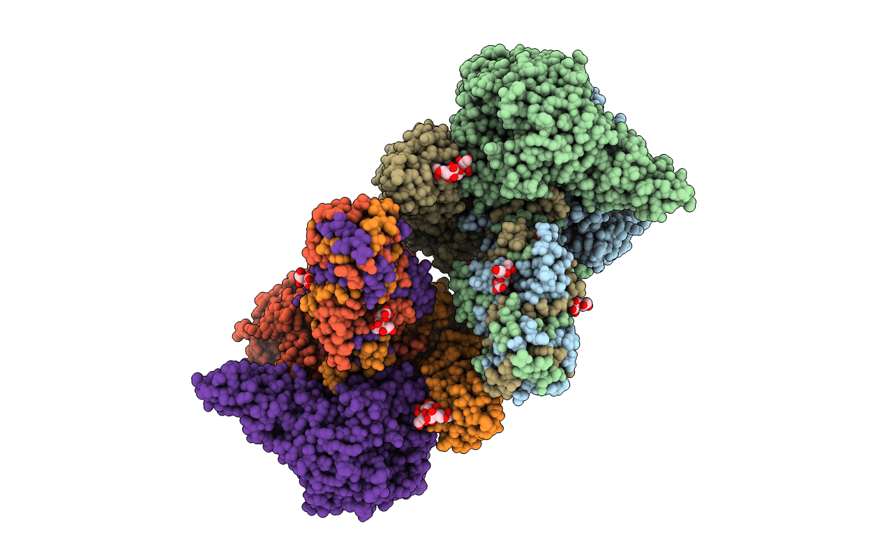
Endosialidase of Bacteriophage K1F in complex with oligomeric alpha-2,8-sialic acid
Biological Source:
Source Organism:
COLIPHAGE K1F (Taxon ID: 344021)
Host Organism:
Method Details:
Experimental Method:
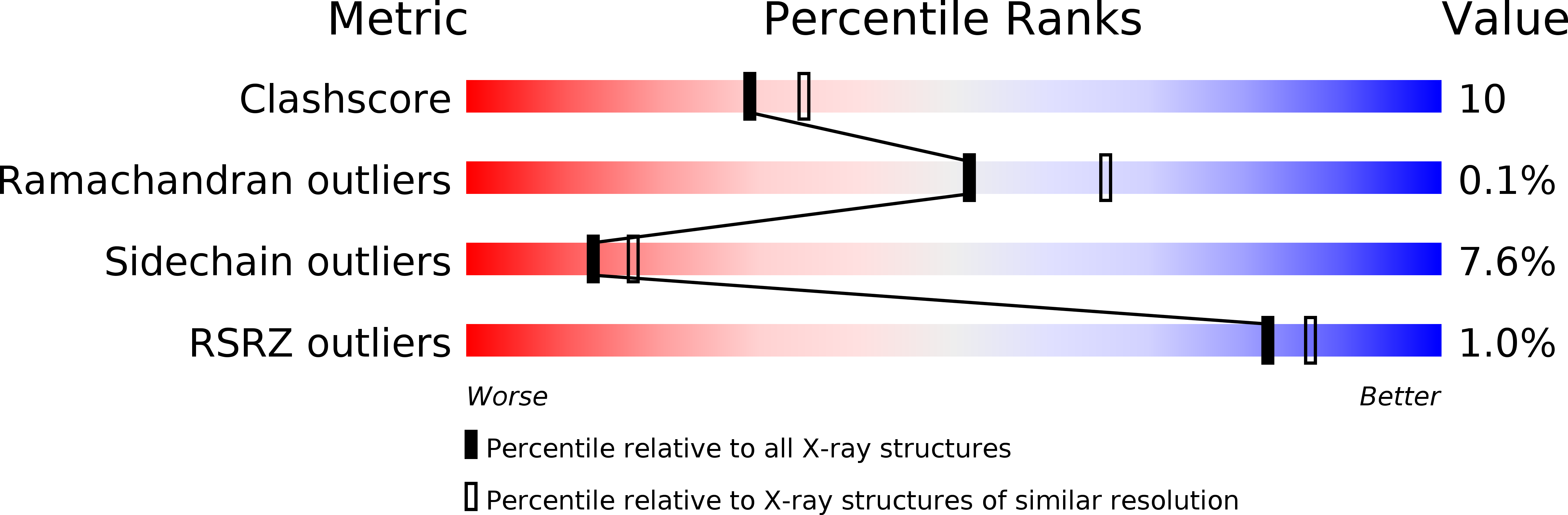
Resolution:
2.55 Å
R-Value Free:
0.23
R-Value Work:
0.18
R-Value Observed:
0.18
Space Group:
P 2 2 21
Macromolecular Entities
Structures with similar UniProt ID
Protein Blast
Polymer Type:polypeptide(L)
Molecule:ENDO-ALPHA-SIALIDASE
Chain IDs:A, B, C, D, E, F
Chain Length:666
Number of Molecules:6
Biological Source:COLIPHAGE K1F
Primary Citation
Crystal Structure of the Polysialic Acid-Degrading Endosialidase of Bacteriophage K1F
Nat.Struct.Mol.Biol. 12 90 ? (2005)
PMID: 15608653 DOI: 10.1038/NSMB874

Abstact

Phages infecting the polysialic acid (polySia)-encapsulated human pathogen Escherichia coli K1 are equipped with capsule-degrading tailspikes known as endosialidases, which are the only identified enzymes that specifically degrade polySia. As polySia also promotes cellular plasticity and tumor metastasis in vertebrates, endosialidases are widely applied in polySia-related neurosciences and cancer research. Here we report the crystal structures of endosialidase NF and its complex with oligomeric sialic acid. The structure NF, which reveals three distinct domains, indicates that the unique polySia specificity evolved from a combination of structural elements characteristic of exosialidases and bacteriophage tailspike proteins. The endosialidase assembles into a catalytic trimer stabilized by a triple beta-helix. Its active site differs markedly from that of exosialidases, indicating an endosialidase-specific substrate-binding mode and catalytic mechanism. Residues essential for endosialidase activity were identified by structure-based mutational analysis.

Legend

Protein

Chemical

Disease

Primary Citation of related structures