Deposition Date
2004-03-22
Release Date
2004-11-17
Last Version Date
2024-05-08
Entry Detail
PDB ID:
1V05
Keywords:
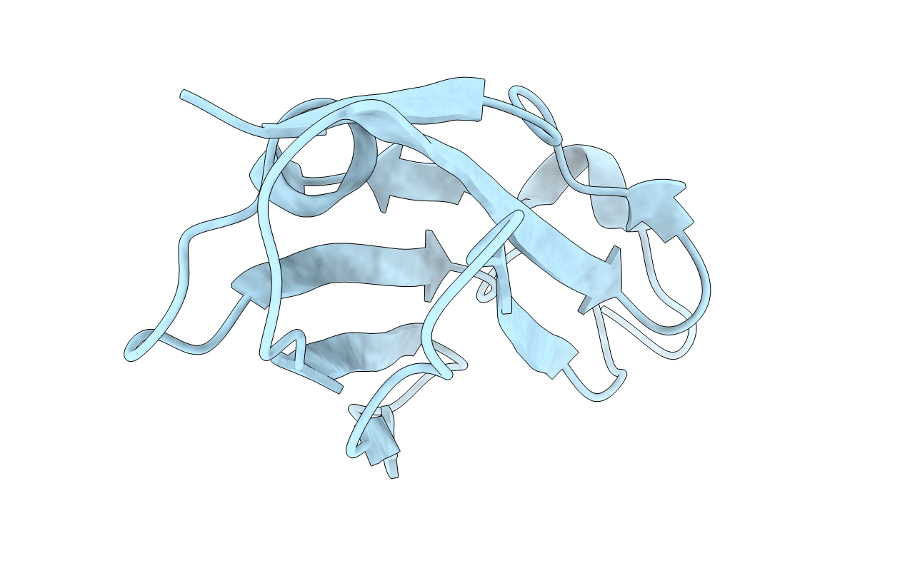
Title:
Dimerization of human Filamin C: crystal structure of the domain 24
Biological Source:
Source Organism(s):
HOMO SAPIENS (Taxon ID: 9606)
Expression System(s):
Method Details:
Experimental Method:
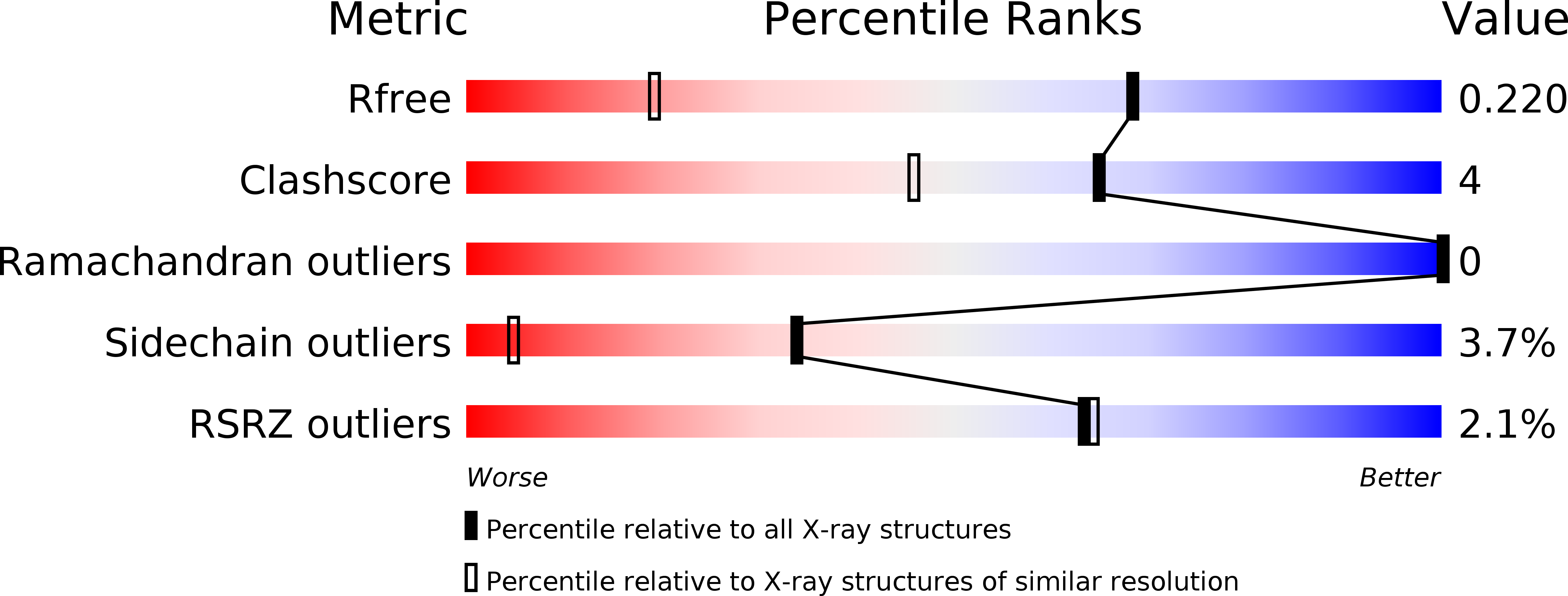
Resolution:
1.43 Å
R-Value Free:
0.20
R-Value Work:
0.18
R-Value Observed:
0.18
Space Group:
P 61 2 2