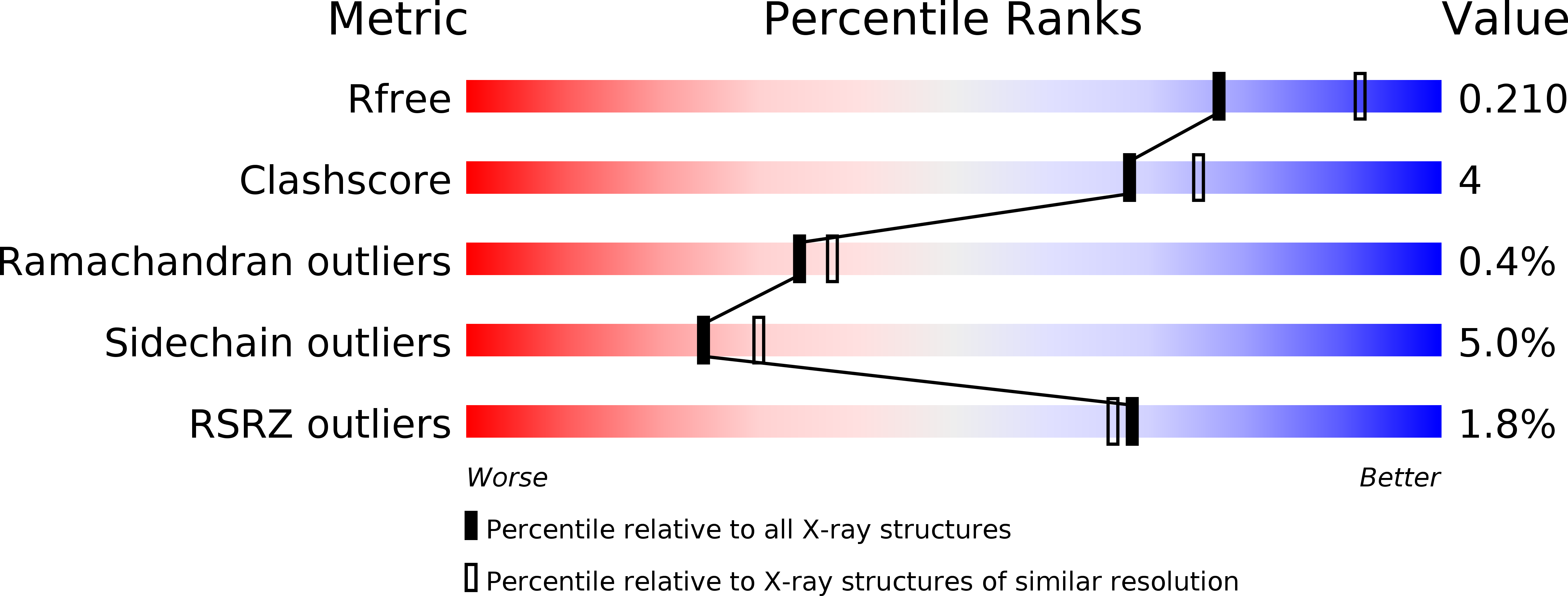
1UZR image
Deposition Date 2004-03-15
Release Date 2004-07-08
Last Version Date 2023-12-13
Entry Detail
PDB ID:
1UZR
Keywords:
Title:
Crystal Structure of the Class Ib Ribonucleotide Reductase R2F-2 subunit from Mycobacterium tuberculosis
Biological Source:
Source Organism:
Host Organism:
Method Details:
Experimental Method:
Resolution:
2.20 Å
R-Value Free:
0.20
R-Value Work:
0.17
R-Value Observed:
0.17
Space Group:
P 42 21 2
Macromolecular Entities
Structures with similar UniProt ID
Protein Blast
Polymer Type:polypeptide(L)
Molecule:RIBONUCLEOTIDE REDUCTASE R2-2 SMALL SUBUNIT
Chain IDs:A, B, C
Chain Length:296
Number of Molecules:3
Biological Source:MYCOBACTERIUM TUBERCULOSIS
Primary Citation
Crystal Structure of the Biologically Active Form of Class Ib Ribonucleotide Reductase Small Subunit from Mycobacterium Tuberculosis
FEBS Lett. 569 117 ? (2004)
PMID: 15225619 DOI: 10.1016/J.FEBSLET.2004.05.059

Abstact

Two nrdF genes of Mycobacterium tuberculosis code for different R2 subunits of the class Ib ribonucleotide reductase (RNR). The proteins are denoted R2F-1 and R2F-2 having 71% sequence identity. The R2F-2 subunit forms the biologically active RNR complex with the catalytic R1E-subunit. We present the structure of the reduced R2F-2 subunit to 2.2 A resolution. Comparison of the R2F-2 structure with a model of R2F-1 suggests that the important differences are located at the C-terminus. We found that within class Ib, the E-helix close to the iron diiron centre has two preferred conformations, which cannot be explained by the redox-state of the diiron centre. In the R2F-2 structure, we also could see a mobility of alphaE in between the two conformations.

Legend

Protein

Chemical

Disease

Primary Citation of related structures