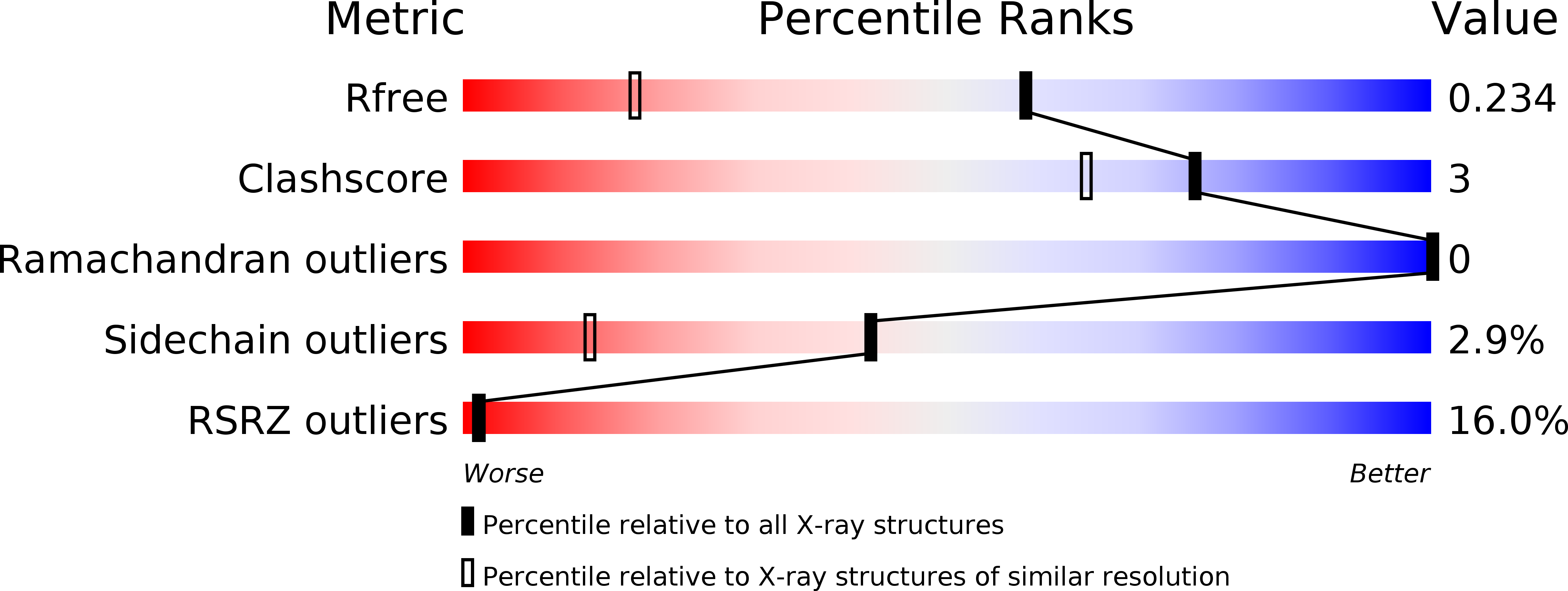
1UZM image
Deposition Date 2004-03-14
Release Date 2005-03-23
Last Version Date 2024-05-08
Entry Detail
PDB ID:
1UZM
Keywords:
Title:
MabA from Mycobacterium tuberculosis
Biological Source:
Source Organism:
Host Organism:
Method Details:
Experimental Method:
Resolution:
1.49 Å
R-Value Free:
0.22
R-Value Work:
0.19
R-Value Observed:
0.19
Space Group:
C 1 2 1
Macromolecular Entities
Polymer Type:polypeptide(L)
Molecule:3-OXOACYL-[ACYL-CARRIER PROTEIN] REDUCTASE
Mutations:YES
Chain IDs:A, B
Chain Length:247
Number of Molecules:2
Biological Source:MYCOBACTERIUM TUBERCULOSIS
Ligand Molecules
Primary Citation
Crystal Structure of Maba from Mycobacterium Tuberculosis, a Reductase Involved in Long-Chain Fatty Acid Biosynthesis.
J.Mol.Biol. 320 249 ? (2002)
PMID: 12079383 DOI: 10.1016/S0022-2836(02)00463-1

Abstact

The fatty acid elongation system FAS-II is involved in the biosynthesis of mycolic acids, which are major and specific long-chain fatty acids of the cell envelope of Mycobacterium tuberculosis and other mycobacteria, including Mycobacterium smegmatis. The protein MabA, also named FabG1, has been shown recently to be part of FAS-II and to catalyse the NADPH-specific reduction of long chain beta-ketoacyl derivatives. This activity corresponds to the second step of an FAS-II elongation round. FAS-II is inhibited by the antituberculous drug isoniazid through the inhibition of the 2-trans-enoyl-acyl carrier protein reductase InhA. Thus, the other enzymes making up this enzymatic complex represent potential targets for designing new antituberculous drugs. The crystal structure of the apo-form MabA was solved to 2.03 A resolution by molecular replacement. MabA is tetrameric and shares the conserved fold of the short-chain dehydrogenases/reductases (SDRs). However, it exhibits some significant local rearrangements of the active-site loops in the absence of a cofactor, particularly the beta5-alpha5 region carrying the unique tryptophan residue, in agreement with previous fluorescence spectroscopy data. A similar conformation has been observed in the beta-ketoacyl reductase from Escherichia coli and the distantly related dehydratase. The distinctive enzymatic and structural properties of MabA are discussed in view of its crystal structure and that of related enzymes.

Legend

Protein

Chemical

Disease

Primary Citation of related structures