Deposition Date
2004-02-20
Release Date
2004-12-07
Last Version Date
2023-12-13
Entry Detail
PDB ID:
1UX8
Keywords:
Title:
X-ray structure of truncated oxygen-avid haemoglobin from Bacillus subtilis
Biological Source:
Source Organism(s):
BACILLUS SUBTILIS (Taxon ID: 1423)
Expression System(s):
Method Details:
Experimental Method:
Resolution:
2.15 Å
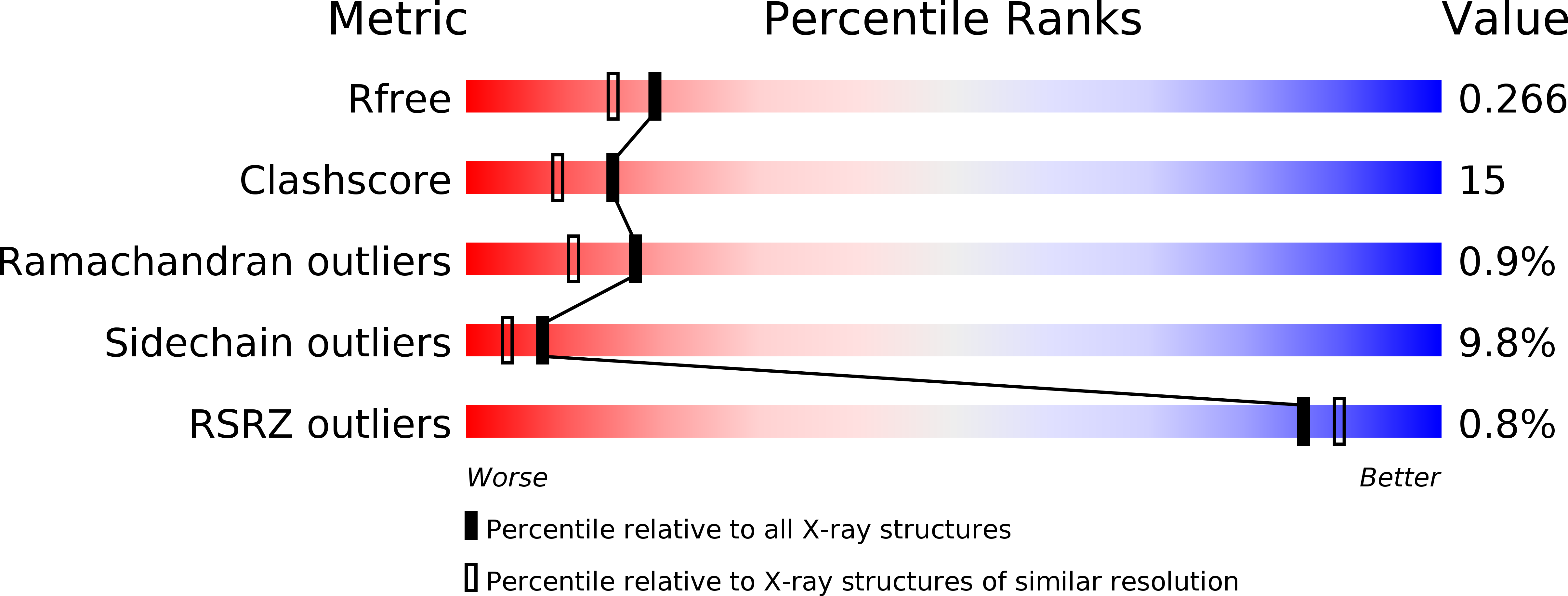
R-Value Free:
0.26
R-Value Work:
0.20
R-Value Observed:
0.20
Space Group:
P 41 21 2