1UWW image
Deposition Date 2004-02-12
Release Date 2004-05-13
Last Version Date 2024-11-06
Entry Detail
PDB ID:
1UWW
Keywords:
Title:
X-ray crystal structure of a non-crystalline cellulose specific carbohydrate-binding module: CBM28.
Biological Source:
Source Organism:
BACILLUS AKIBAI (Taxon ID: 1411)
Host Organism:
Method Details:
Experimental Method:
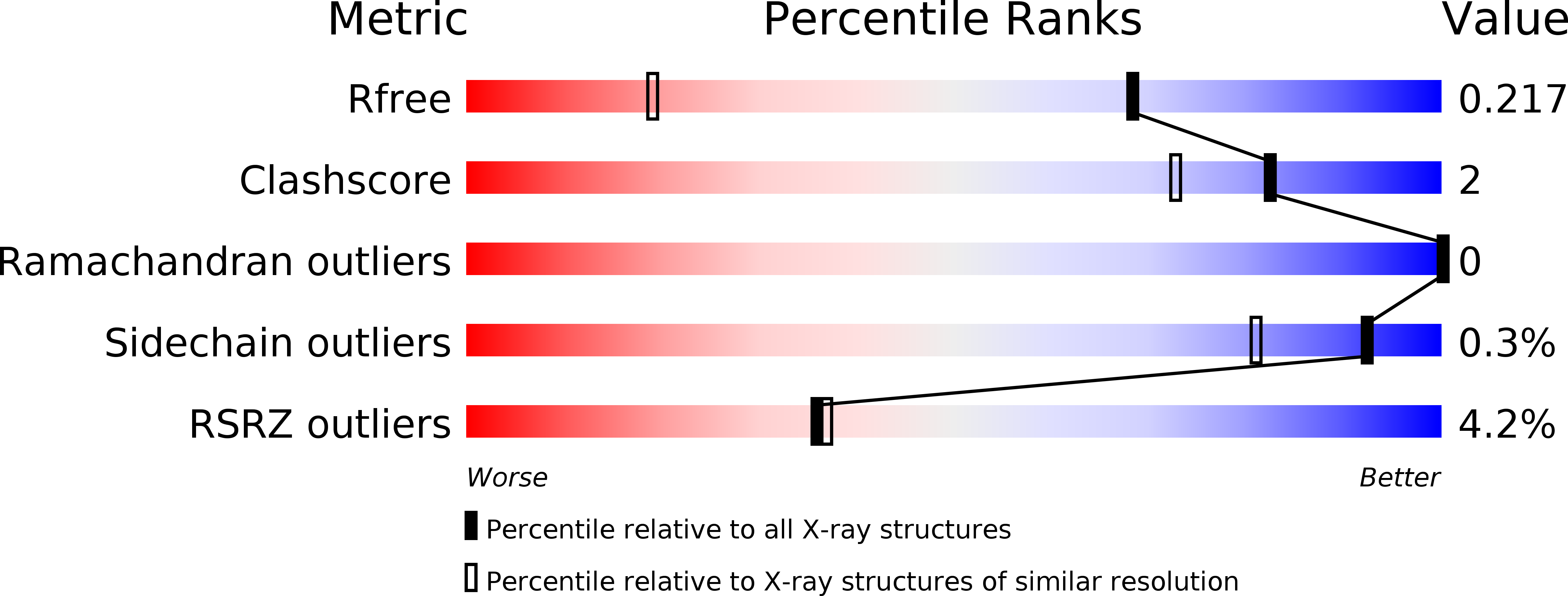
Resolution:
1.40 Å
R-Value Free:
0.20
R-Value Work:
0.18
R-Value Observed:
0.19
Space Group:
P 21 21 21
Macromolecular Entities
Structures with similar UniProt ID
Protein Blast
Polymer Type:polypeptide(L)
Molecule:ENDOGLUCANASE
Chain IDs:A, B
Chain Length:191
Number of Molecules:2
Biological Source:BACILLUS AKIBAI
Modified Residue
Compound ID Chain ID Parent Comp ID Details 2D Image
MSE A MET SELENOMETHIONINE
Ligand Molecules
Primary Citation
X-Ray Crystal Structure of a Non-Crystalline Cellulose-Specific Carbohydrate-Binding Module: Cbm28
J.Mol.Biol. 339 253 ? (2004)
PMID: 15136030 DOI: 10.1016/J.JMB.2004.03.069

Abstact

Natural cellulose exists as a composite of different forms, which have historically been broadly characterized as "crystalline" or "amorphous". The recognition of both of these forms of cellulose by the carbohydrate-binding modules (CBM) of microbial glycoside hydrolases is central to natural and efficient biotechnological conversion of plant cell wall biomass. There is increasing evidence that, at least some, individual binding modules target distinct and different regions of non-crystalline "amorphous" cellulose. Competition experiments show that CBM28 modules do not compete with CBM17 modules when binding to non-crystalline cellulose. The structure of the BspCBM28 (http://afmb.cnrs-mrs.fr/CAZY/) module from the Bacillus sp. 1139 family GH5 endoglucanase, comprising a 191 amino acid protein, has therefore been determined at 1.4A resolution using single isomorphous replacement with anomalous scattering methods. The structure reveals a "beta-jelly roll" topology, with high degree of similarity to the structure of CBM17 domains. Sequence and structural conservation strongly suggests that these two families of domains have evolved through gene duplication and subsequent divergence. The ligand-binding site "topographies" of CBMs from families 28, 17 and 4 begins to shed light on the differential recognition of non-crystalline cellulose by multi-modular plant cell wall-degrading enzymes.

Legend

Protein

Chemical

Disease

Primary Citation of related structures